 Movie
Movie Controller


+ Open data

- Basic information
| Entry | Database: PDB / ID: 1r1g | ||||||
|---|---|---|---|---|---|---|---|
| Title | Crystal Structure of the Scorpion Toxin BmBKTtx1 | ||||||
 Components Components | Neurotoxin BmK37 | ||||||
 Keywords Keywords | TOXIN / SIRAS from S atoms / scorpion toxin / BmBKTtx1 / reductive dimethylation | ||||||
| Function / homology | Scorpion short toxins signature. / Scorpion short chain toxin, potassium channel inhibitor / Scorpion short toxin, BmKK2 / Knottin, scorpion toxin-like superfamily / ion channel inhibitor activity / potassium channel regulator activity / toxin activity / extracellular region / Potassium channel toxin alpha-KTx 19.1 Function and homology information Function and homology information | ||||||
| Method |  X-RAY DIFFRACTION / SIRAS / Resolution: 1.72 Å X-RAY DIFFRACTION / SIRAS / Resolution: 1.72 Å | ||||||
 Authors Authors | Szyk, A. / Lu, W. / Xu, C. / Lubkowski, J. | ||||||
 Citation Citation | Journal: J.Struct.Biol. / Year: 2004 Title: Structure of the scorpion toxin BmBKTtx1 solved from single wavelength anomalous scattering of sulfur. Authors: Szyk, A. / Lu, W. / Xu, C. / Lubkowski, J. | ||||||
| History |
| ||||||
| Remark 999 | SEQUENCE ALL PRIMARY AMINO GROUPS (FIVE LYSINE RESIDUES AND THE N-TERMINUS) WERE DIMETHYLATED. ...SEQUENCE ALL PRIMARY AMINO GROUPS (FIVE LYSINE RESIDUES AND THE N-TERMINUS) WERE DIMETHYLATED. DENSITY WAS NOT SUFFICIENT TO MODEL THE METHYL GROUPS FOR THE N-TERMINUS. |
- Structure visualization
| Structure viewer | Molecule: MolmilJmol/JSmol |
|---|
- Downloads & links
-Download
| PDBx/mmCIF format | 1r1g.cif.gz | 26.1 KB | Display | PDBx/mmCIF format |
|---|---|---|---|---|
| PDB format | pdb1r1g.ent.gz | 17.4 KB | Display | PDB format |
| PDBx/mmJSON format | 1r1g.json.gz | Tree view | PDBx/mmJSON format | |
| Others |  Other downloads Other downloads |
-Validation report
| Arichive directory | https://data.pdbj.org/pub/pdb/validation_reports/r1/1r1gftp://data.pdbj.org/pub/pdb/validation_reports/r1/1r1g | HTTPS FTP |
|---|
-Related structure data
| Related structure data | |
|---|---|
| Similar structure data |

-Links
 PDBj
PDBj

- Assembly
| Deposited unit | 
| ||||||||
|---|---|---|---|---|---|---|---|---|---|
| 1 | 
| ||||||||
| 2 | 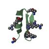
| ||||||||
| Unit cell |
| ||||||||
| Details | Biologically active form of BmBKTx1 is a monomer. |
-Components
| #1: Protein/peptide | Mass: 3511.301 Da / Num. of mol.: 2 / Source method: obtained synthetically Details: Sequence corresponds to that of naturally occuring toxin from scorpion Buthus martensi Karsch. Protein was synthesized using Boc chemistry. After folding all primary amino groups of BmBKTtx1 ...Details: Sequence corresponds to that of naturally occuring toxin from scorpion Buthus martensi Karsch. Protein was synthesized using Boc chemistry. After folding all primary amino groups of BmBKTtx1 (five lysine residues and the N-terminus) were dimethylated. References: UniProt: P83407 #2: Water | ChemComp-HOH / |  Mass: 18.015 Da / Num. of mol.: 108 / Source method: isolated from a natural source / Formula: H2O Mass: 18.015 Da / Num. of mol.: 108 / Source method: isolated from a natural source / Formula: H2OHas protein modification | Y | |
|---|
-Experimental details
-Experiment
| Experiment | Method: X-RAY DIFFRACTION / Number of used crystals: 1 |
|---|
- Sample preparation
| Crystal | Density Matthews: 1.77 Å3/Da / Density % sol: 30.56 % |
|---|---|
| Crystal grow | Temperature: 293 K / Method: vapor diffusion, hanging drop Details: Sodium Formate, VAPOR DIFFUSION, HANGING DROP, temperature 293.0K |
| Crystal grow | *PLUS Method: sparse-matrix |
| Components of the solutions | *PLUS Conc.: 100 mg/ml / Common name: protein |
-Data collection
| Diffraction | Mean temperature: 100 K |
|---|---|
| Diffraction source | Source: ROTATING ANODE / Type: RIGAKU RU200 / Wavelength: 1.5478 Å |
| Detector | Type: MARRESEARCH / Detector: IMAGE PLATE / Date: Aug 15, 2003 / Details: Osmic mirrors |
| Radiation | Protocol: SINGLE WAVELENGTH / Monochromatic (M) / Laue (L): M / Scattering type: x-ray |
| Radiation wavelength | Wavelength: 1.5478 Å / Relative weight: 1 |
| Reflection | Resolution: 1.72→30 Å / Num. all: 5064 / Num. obs: 5028 / % possible obs: 95.4 % / Observed criterion σ(I): 1 / Redundancy: 12.6 % / Biso Wilson estimate: 14.3 Å2 / Rmerge(I) obs: 0.036 / Rsym value: 0.036 / Net I/σ(I): 65.5 |
| Reflection shell | Resolution: 1.72→1.78 Å / Redundancy: 12.3 % / Rmerge(I) obs: 0.089 / Mean I/σ(I) obs: 19 / Num. unique all: 491 / Rsym value: 0.089 / % possible all: 91.6 |
| Reflection | *PLUS Num. obs: 5064 / Num. measured all: 9834 |
| Reflection shell | *PLUS % possible obs: 91.6 % |
- Processing
| Software |
| ||||||||||||||||||||||||||||||||||||
|---|---|---|---|---|---|---|---|---|---|---|---|---|---|---|---|---|---|---|---|---|---|---|---|---|---|---|---|---|---|---|---|---|---|---|---|---|---|
| Refinement | Method to determine structure: SIRAS / Resolution: 1.72→14.65 Å / Rfactor Rfree error: 0.014 / Isotropic thermal model: RESTRAINED / Cross valid method: THROUGHOUT / σ(F): 0
| ||||||||||||||||||||||||||||||||||||
| Solvent computation | Solvent model: FLAT MODEL / Bsol: 77.9579 Å2 / ksol: 0.428433 e/Å3 | ||||||||||||||||||||||||||||||||||||
| Displacement parameters | Biso mean: 17.4 Å2
| ||||||||||||||||||||||||||||||||||||
| Refine analyze |
| ||||||||||||||||||||||||||||||||||||
| Refinement step | Cycle: LAST / Resolution: 1.72→14.65 Å
| ||||||||||||||||||||||||||||||||||||
| Refine LS restraints |
| ||||||||||||||||||||||||||||||||||||
| LS refinement shell | Resolution: 1.72→1.83 Å / Rfactor Rfree error: 0.032 / Total num. of bins used: 6
| ||||||||||||||||||||||||||||||||||||
| Xplor file |
| ||||||||||||||||||||||||||||||||||||
| Refinement | *PLUS Lowest resolution: 15 Å / Num. reflection obs: 4756 / % reflection Rfree: 5 % | ||||||||||||||||||||||||||||||||||||
| Solvent computation | *PLUS | ||||||||||||||||||||||||||||||||||||
| Displacement parameters | *PLUS | ||||||||||||||||||||||||||||||||||||
| Refine LS restraints | *PLUS
|
