 Movie
Movie Controller


+ Open data

- Basic information
| Entry | Database: PDB / ID: 1jbb | ||||||
|---|---|---|---|---|---|---|---|
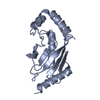
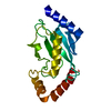
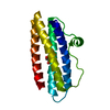
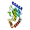
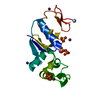
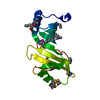
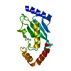
| Title | Ubiquitin Conjugating Enzyme, Ubc13 | ||||||
 Components Components | ubiquitin conjugating enzyme E2-17.5 KDA | ||||||
 Keywords Keywords | LIGASE / UBIQUITIN / UBIQUITIN-CONJUGATING ENZYME / E2 | ||||||
| Function / homology |  Function and homology information Function and homology informationprotein targeting to vacuolar membrane / : / Aggrephagy / PINK1-PRKN Mediated Mitophagy / free ubiquitin chain polymerization / ubiquitin conjugating enzyme complex / : / Recruitment and ATM-mediated phosphorylation of repair and signaling proteins at DNA double strand breaks / fungal-type vacuole membrane / DNA damage tolerance ...protein targeting to vacuolar membrane / : / Aggrephagy / PINK1-PRKN Mediated Mitophagy / free ubiquitin chain polymerization / ubiquitin conjugating enzyme complex / : / Recruitment and ATM-mediated phosphorylation of repair and signaling proteins at DNA double strand breaks / fungal-type vacuole membrane / DNA damage tolerance / E2 ubiquitin-conjugating enzyme / ubiquitin conjugating enzyme activity / Antigen processing: Ubiquitination & Proteasome degradation / protein K63-linked ubiquitination / protein polyubiquitination / ATP binding / nucleus / cytoplasm / cytosol Similarity search - Function | ||||||
| Biological species |  | ||||||
| Method |  X-RAY DIFFRACTION / MOLECULAR REPLACEMENT / Resolution: 2 Å X-RAY DIFFRACTION / MOLECULAR REPLACEMENT / Resolution: 2 Å | ||||||
 Authors Authors | VanDemark, A.P. / Hofmann, R.M. / Tsui, C. / Pickart, C.M. / Wolberger, C. | ||||||
 Citation Citation | Journal: Cell(Cambridge,Mass.) / Year: 2001 Title: Molecular insights into polyubiquitin chain assembly: crystal structure of the Mms2/Ubc13 heterodimer. Authors: VanDemark, A.P. / Hofmann, R.M. / Tsui, C. / Pickart, C.M. / Wolberger, C. | ||||||
| History |
|
- Structure visualization
| Structure viewer | Molecule: MolmilJmol/JSmol |
|---|
- Downloads & links
-Download
| PDBx/mmCIF format | 1jbb.cif.gz | 71.5 KB | Display | PDBx/mmCIF format |
|---|---|---|---|---|
| PDB format | pdb1jbb.ent.gz | 53.9 KB | Display | PDB format |
| PDBx/mmJSON format | 1jbb.json.gz | Tree view | PDBx/mmJSON format | |
| Others |  Other downloads Other downloads |
-Validation report
| Arichive directory | https://data.pdbj.org/pub/pdb/validation_reports/jb/1jbbftp://data.pdbj.org/pub/pdb/validation_reports/jb/1jbb | HTTPS FTP |
|---|
-Related structure data
| Related structure data | 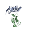 1jatC 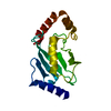 1qcqS S: Starting model for refinement C: citing same article ( |
|---|---|
| Similar structure data |


-Links
 PDBj
PDBj


- Assembly
| Deposited unit | 
| ||||||||
|---|---|---|---|---|---|---|---|---|---|
| 1 | 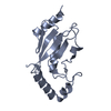
| ||||||||
| 2 | 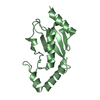
| ||||||||
| Unit cell |
|
-Components
| #1: Protein | Mass: 17489.984 Da / Num. of mol.: 2 Source method: isolated from a genetically manipulated source Source: (gene. exp.) Description: GST-fusion / Gene: Ubc13 / Plasmid: pGEX / Production host:  #2: Water | ChemComp-HOH / |  Mass: 18.015 Da / Num. of mol.: 131 / Source method: isolated from a natural source / Formula: H2O Mass: 18.015 Da / Num. of mol.: 131 / Source method: isolated from a natural source / Formula: H2O |
|---|
-Experimental details
-Experiment
| Experiment | Method: X-RAY DIFFRACTION / Number of used crystals: 1 |
|---|
- Sample preparation
| Crystal | Density Matthews: 2.05 Å3/Da / Density % sol: 39.88 % | ||||||||||||||||||||||||||||||||||||||||||||||||||||||
|---|---|---|---|---|---|---|---|---|---|---|---|---|---|---|---|---|---|---|---|---|---|---|---|---|---|---|---|---|---|---|---|---|---|---|---|---|---|---|---|---|---|---|---|---|---|---|---|---|---|---|---|---|---|---|---|
| Crystal grow | Temperature: 298 K / Method: vapor diffusion, hanging drop / pH: 6 Details: PEG 5000-MME, ammonium sulfate, pH 6.0, VAPOR DIFFUSION, HANGING DROP, temperature 298K | ||||||||||||||||||||||||||||||||||||||||||||||||||||||
| Crystal grow | *PLUS Temperature: 20 ℃ / pH: 8 | ||||||||||||||||||||||||||||||||||||||||||||||||||||||
| Components of the solutions | *PLUS
|
-Data collection
| Diffraction | Mean temperature: 298 K |
|---|---|
| Diffraction source | Source: ROTATING ANODE / Type: RIGAKU RU200 / Wavelength: 1.542 Å |
| Detector | Type: RIGAKU RAXIS IV / Detector: IMAGE PLATE / Date: Jul 30, 2000 / Details: mirrors |
| Radiation | Monochromator: graphite / Protocol: SINGLE WAVELENGTH / Monochromatic (M) / Laue (L): M / Scattering type: x-ray |
| Radiation wavelength | Wavelength: 1.542 Å / Relative weight: 1 |
| Reflection | Resolution: 2→50 Å / Num. all: 17787 / Num. obs: 17787 / % possible obs: 93.6 % / Observed criterion σ(F): 0 / Observed criterion σ(I): 0 / Redundancy: 3.4 % / Biso Wilson estimate: 20.099 Å2 / Rmerge(I) obs: 0.053 / Net I/σ(I): 27.8 |
| Reflection shell | Resolution: 2→2.09 Å / Rmerge(I) obs: 0.223 / Mean I/σ(I) obs: 4.4 / Num. unique all: 1937 / % possible all: 83.2 |
| Reflection | *PLUS Lowest resolution: 50 Å / Num. measured all: 55421 |
| Reflection shell | *PLUS % possible obs: 83.2 % |
- Processing
| Software |
| ||||||||||||||||||||
|---|---|---|---|---|---|---|---|---|---|---|---|---|---|---|---|---|---|---|---|---|---|
| Refinement | Method to determine structure: MOLECULAR REPLACEMENT Starting model: PDB entry 1QCQ truncated to ALAs Resolution: 2→40 Å / Isotropic thermal model: anisotropic / Cross valid method: THROUGHOUT / σ(F): 0 / σ(I): 0 / Stereochemistry target values: Engh & Huber
| ||||||||||||||||||||
| Refine analyze |
| ||||||||||||||||||||
| Refinement step | Cycle: LAST / Resolution: 2→40 Å
| ||||||||||||||||||||
| Refine LS restraints |
| ||||||||||||||||||||
| LS refinement shell | Resolution: 2→2.09 Å
| ||||||||||||||||||||
| Software | *PLUS Name: CNS / Classification: refinement | ||||||||||||||||||||
| Refinement | *PLUS Highest resolution: 2 Å / Lowest resolution: 40 Å / σ(F): 0 / % reflection Rfree: 5 % / Rfactor obs: 0.188 | ||||||||||||||||||||
| Solvent computation | *PLUS | ||||||||||||||||||||
| Displacement parameters | *PLUS | ||||||||||||||||||||
| LS refinement shell | *PLUS Rfactor Rfree: 0.268 |
