 Movie
Movie Controller


+ Open data

- Basic information
| Entry | Database: PDB / ID: 7bol | ||||||
|---|---|---|---|---|---|---|---|
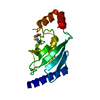
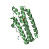
| Title | ubiquitin-conjugating enzyme, Ube2D2 | ||||||
 Components Components | Ubiquitin-conjugating enzyme E2 D2 | ||||||
 Keywords Keywords | STRUCTURAL PROTEIN / ubiquitin-conjugating enzyme / Ube2D2 | ||||||
| Function / homology |  Function and homology information Function and homology information(E3-independent) E2 ubiquitin-conjugating enzyme / E2 ubiquitin-conjugating enzyme / ubiquitin conjugating enzyme activity / protein autoubiquitination / protein K48-linked ubiquitination / protein modification process / Synthesis of active ubiquitin: roles of E1 and E2 enzymes / TICAM1, RIP1-mediated IKK complex recruitment / IKK complex recruitment mediated by RIP1 / PINK1-PRKN Mediated Mitophagy ...(E3-independent) E2 ubiquitin-conjugating enzyme / E2 ubiquitin-conjugating enzyme / ubiquitin conjugating enzyme activity / protein autoubiquitination / protein K48-linked ubiquitination / protein modification process / Synthesis of active ubiquitin: roles of E1 and E2 enzymes / TICAM1, RIP1-mediated IKK complex recruitment / IKK complex recruitment mediated by RIP1 / PINK1-PRKN Mediated Mitophagy / Negative regulators of DDX58/IFIH1 signaling / Peroxisomal protein import / Regulation of TNFR1 signaling / Inactivation of CSF3 (G-CSF) signaling / Oxygen-dependent proline hydroxylation of Hypoxia-inducible Factor Alpha / CLEC7A (Dectin-1) signaling / FCERI mediated NF-kB activation / protein polyubiquitination / ubiquitin-protein transferase activity / ubiquitin protein ligase activity / Downstream TCR signaling / Antigen processing: Ubiquitination & Proteasome degradation / E3 ubiquitin ligases ubiquitinate target proteins / Neddylation / ubiquitin-dependent protein catabolic process / protein ubiquitination / protein-containing complex / extracellular exosome / nucleoplasm / ATP binding / nucleus / cytosol Similarity search - Function | ||||||
| Biological species |  Homo sapiens (human) Homo sapiens (human) | ||||||
| Method |  X-RAY DIFFRACTION / SYNCHROTRON / MOLECULAR REPLACEMENT / Resolution: 1.797 Å X-RAY DIFFRACTION / SYNCHROTRON / MOLECULAR REPLACEMENT / Resolution: 1.797 Å | ||||||
 Authors Authors | Lee, S.O. / Ryu, K.S. / Chi, S.-W. | ||||||
 Citation Citation | Journal: Febs J. / Year: 2022 Title: MUL1-RING recruits the substrate, p53-TAD as a complex with UBE2D2-UB conjugate. Authors: Lee, M.S. / Lee, S.O. / Choi, J. / Ryu, M. / Lee, M.K. / Kim, J.H. / Hwang, E. / Lee, C.K. / Chi, S.W. / Ryu, K.S. | ||||||
| History |
|
- Structure visualization
| Structure viewer | Molecule: MolmilJmol/JSmol |
|---|
- Downloads & links
-Download
| PDBx/mmCIF format | 7bol.cif.gz | 69.2 KB | Display | PDBx/mmCIF format |
|---|---|---|---|---|
| PDB format | pdb7bol.ent.gz | 50.8 KB | Display | PDB format |
| PDBx/mmJSON format | 7bol.json.gz | Tree view | PDBx/mmJSON format | |
| Others |  Other downloads Other downloads |
-Validation report
| Arichive directory | https://data.pdbj.org/pub/pdb/validation_reports/bo/7bolftp://data.pdbj.org/pub/pdb/validation_reports/bo/7bol | HTTPS FTP |
|---|
-Related structure data
| Related structure data |  6m2cC  6m2dC  2eskS S: Starting model for refinement C: citing same article ( |
|---|---|
| Similar structure data |


-Links
 PDBj
PDBj


- Assembly
| Deposited unit | 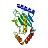
| ||||||||
|---|---|---|---|---|---|---|---|---|---|
| 1 |
| ||||||||
| Unit cell |
|
-Components
| #1: Protein | Mass: 16842.305 Da / Num. of mol.: 1 Source method: isolated from a genetically manipulated source Source: (gene. exp.) Homo sapiens (human) / Gene: UBE2D2, PUBC1, UBC4, UBC5B, UBCH4, UBCH5B / Production host:  References: UniProt: P62837, E2 ubiquitin-conjugating enzyme, (E3-independent) E2 ubiquitin-conjugating enzyme |
|---|---|
| #2: Water | ChemComp-HOH /  Mass: 18.015 Da / Num. of mol.: 124 / Source method: isolated from a natural source / Formula: H2O Mass: 18.015 Da / Num. of mol.: 124 / Source method: isolated from a natural source / Formula: H2O |
-Experimental details
-Experiment
| Experiment | Method: X-RAY DIFFRACTION / Number of used crystals: 1 |
|---|
- Sample preparation
| Crystal | Density Matthews: 2.37 Å3/Da / Density % sol: 48.12 % |
|---|---|
| Crystal grow | Temperature: 291 K / Method: vapor diffusion, sitting drop Details: 0.5 M Ammonium sulfate 0.1 M Sodium citrate pH 5.6 1.0 M Lithium sulfate |
-Data collection
| Diffraction | Mean temperature: 93 K / Serial crystal experiment: N |
|---|---|
| Diffraction source | Source: SYNCHROTRON / Site: PAL/PLS  / Beamline: 7A (6B, 6C1) / Wavelength: 0.9793 Å / Beamline: 7A (6B, 6C1) / Wavelength: 0.9793 Å |
| Detector | Type: ADSC QUANTUM 270 / Detector: CCD / Date: May 4, 2016 |
| Radiation | Protocol: MAD / Monochromatic (M) / Laue (L): M / Scattering type: x-ray |
| Radiation wavelength | Wavelength: 0.9793 Å / Relative weight: 1 |
| Reflection | Resolution: 1.797→50 Å / Num. obs: 14967 / % possible obs: 96.8 % / Redundancy: 6.1 % / Biso Wilson estimate: 15.47 Å2 / Rmerge(I) obs: 0.072 / Net I/σ(I): 26.3 |
- Processing
| Software |
| ||||||||||||||||||||||||||||||||||||
|---|---|---|---|---|---|---|---|---|---|---|---|---|---|---|---|---|---|---|---|---|---|---|---|---|---|---|---|---|---|---|---|---|---|---|---|---|---|
| Refinement | Method to determine structure: MOLECULAR REPLACEMENT Starting model: 2ESK Resolution: 1.797→26.476 Å / SU ML: 0.23 / Cross valid method: THROUGHOUT / σ(F): 1.5 / Phase error: 20.4
| ||||||||||||||||||||||||||||||||||||
| Solvent computation | Shrinkage radii: 0.9 Å / VDW probe radii: 1.11 Å | ||||||||||||||||||||||||||||||||||||
| Displacement parameters | Biso max: 63.25 Å2 / Biso mean: 18.3873 Å2 / Biso min: 5.22 Å2 | ||||||||||||||||||||||||||||||||||||
| Refinement step | Cycle: final / Resolution: 1.797→26.476 Å
| ||||||||||||||||||||||||||||||||||||
| Refine LS restraints |
| ||||||||||||||||||||||||||||||||||||
| LS refinement shell | Refine-ID: X-RAY DIFFRACTION / Rfactor Rfree error: 0
|
