 Movie
Movie Controller


+ Open data

- Basic information
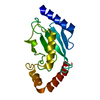
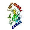
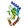
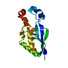
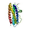
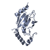
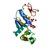
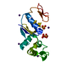
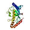
| Entry | Database: PDB / ID: 2c4p | ||||||
|---|---|---|---|---|---|---|---|
| Title | Crystal structure of human ubiquitin-conjugating enzyme UbcH5A | ||||||
 Components Components | UBIQUITIN-CONJUGATING ENZYME E2 D1 | ||||||
 Keywords Keywords | LIGASE / THIOESTERIFICATION / UBIQUITIN-CONJUGATING ENZYME / UBL CONJUGATION PATHWAY | ||||||
| Function / homology |  Function and homology information Function and homology informationpositive regulation of protein polyubiquitination / Conversion from APC/C:Cdc20 to APC/C:Cdh1 in late anaphase / Inactivation of APC/C via direct inhibition of the APC/C complex / APC/C:Cdc20 mediated degradation of mitotic proteins / Aberrant regulation of mitotic exit in cancer due to RB1 defects / Phosphorylation of the APC/C / Signaling by BMP / (E3-independent) E2 ubiquitin-conjugating enzyme / E2 ubiquitin-conjugating enzyme / Regulation of APC/C activators between G1/S and early anaphase ...positive regulation of protein polyubiquitination / Conversion from APC/C:Cdc20 to APC/C:Cdh1 in late anaphase / Inactivation of APC/C via direct inhibition of the APC/C complex / APC/C:Cdc20 mediated degradation of mitotic proteins / Aberrant regulation of mitotic exit in cancer due to RB1 defects / Phosphorylation of the APC/C / Signaling by BMP / (E3-independent) E2 ubiquitin-conjugating enzyme / E2 ubiquitin-conjugating enzyme / Regulation of APC/C activators between G1/S and early anaphase / ubiquitin conjugating enzyme activity / Transcriptional Regulation by VENTX / negative regulation of BMP signaling pathway / negative regulation of TORC1 signaling / protein K48-linked ubiquitination / APC/C:Cdc20 mediated degradation of Cyclin B / APC-Cdc20 mediated degradation of Nek2A / positive regulation of protein ubiquitination / Synthesis of active ubiquitin: roles of E1 and E2 enzymes / TICAM1, RIP1-mediated IKK complex recruitment / IKK complex recruitment mediated by RIP1 / Autodegradation of Cdh1 by Cdh1:APC/C / APC/C:Cdc20 mediated degradation of Securin / Negative regulators of DDX58/IFIH1 signaling / Peroxisomal protein import / Downregulation of SMAD2/3:SMAD4 transcriptional activity / Assembly of the pre-replicative complex / Regulation of TNFR1 signaling / Cdc20:Phospho-APC/C mediated degradation of Cyclin A / Degradation of CRY and PER proteins / Inactivation of CSF3 (G-CSF) signaling / APC/C:Cdh1 mediated degradation of Cdc20 and other APC/C:Cdh1 targeted proteins in late mitosis/early G1 / Oxygen-dependent proline hydroxylation of Hypoxia-inducible Factor Alpha / CDK-mediated phosphorylation and removal of Cdc6 / CLEC7A (Dectin-1) signaling / FCERI mediated NF-kB activation / protein polyubiquitination / ubiquitin-protein transferase activity / Ovarian tumor domain proteases / Separation of Sister Chromatids / ubiquitin protein ligase activity / Downstream TCR signaling / Antigen processing: Ubiquitination & Proteasome degradation / E3 ubiquitin ligases ubiquitinate target proteins / Neddylation / Senescence-Associated Secretory Phenotype (SASP) / ubiquitin-dependent protein catabolic process / proteasome-mediated ubiquitin-dependent protein catabolic process / negative regulation of transcription by RNA polymerase II / protein-containing complex / nucleoplasm / ATP binding / nucleus / cytoplasm / cytosol Similarity search - Function | ||||||
| Biological species |  HOMO SAPIENS (human) HOMO SAPIENS (human) | ||||||
| Method |  X-RAY DIFFRACTION / SYNCHROTRON / MOLECULAR REPLACEMENT / Resolution: 2.35 Å X-RAY DIFFRACTION / SYNCHROTRON / MOLECULAR REPLACEMENT / Resolution: 2.35 Å | ||||||
 Authors Authors | Dodd, R.B. / Read, R.J. | ||||||
 Citation Citation | Journal: To be Published Title: Structures of Two Human Ubiquitin-Conjugating Enzymes from Twinned Crystals Authors: Dodd, R.B. / Read, R.J. | ||||||
| History |
|
- Structure visualization
| Structure viewer | Molecule: MolmilJmol/JSmol |
|---|
- Downloads & links
-Download
| PDBx/mmCIF format | 2c4p.cif.gz | 73.1 KB | Display | PDBx/mmCIF format |
|---|---|---|---|---|
| PDB format | pdb2c4p.ent.gz | 54.3 KB | Display | PDB format |
| PDBx/mmJSON format | 2c4p.json.gz | Tree view | PDBx/mmJSON format | |
| Others |  Other downloads Other downloads |
-Validation report
| Arichive directory | https://data.pdbj.org/pub/pdb/validation_reports/c4/2c4pftp://data.pdbj.org/pub/pdb/validation_reports/c4/2c4p | HTTPS FTP |
|---|
-Related structure data
| Related structure data |  2clwC 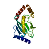 1qcqS S: Starting model for refinement C: citing same article ( |
|---|---|
| Similar structure data |

-Links
 PDBj
PDBj


- Assembly
| Deposited unit | 
| ||||||||
|---|---|---|---|---|---|---|---|---|---|
| 1 | 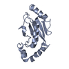
| ||||||||
| 2 | 
| ||||||||
| Unit cell |
| ||||||||
| Noncrystallographic symmetry (NCS) | NCS oper: (Code: given Matrix: (0.99647, -0.08301, 0.01224), Vector: |
-Components
| #1: Protein | Mass: 18694.447 Da / Num. of mol.: 2 Source method: isolated from a genetically manipulated source Source: (gene. exp.) HOMO SAPIENS (human) / Plasmid: PETM-12 (EMBL) / Production host:  #2: Water | ChemComp-HOH / |  Mass: 18.015 Da / Num. of mol.: 115 / Source method: isolated from a natural source / Formula: H2O Mass: 18.015 Da / Num. of mol.: 115 / Source method: isolated from a natural source / Formula: H2OCompound details | CATALYZES THE COVALENT ATTACHMENT OF UBIQUITIN TO OTHER PROTEINS. MEDIATES THE SELECTIVE ...CATALYZES THE COVALENT ATTACHMENT | Sequence details | RESIDUES FOR WHICH INSUFFICIENT DENSITY WAS OBSERVED FOR SIDECHAINS WERE TRUNCATED TO ALANINES. THE ...RESIDUES FOR WHICH INSUFFICIE | |
|---|
-Experimental details
-Experiment
| Experiment | Method: X-RAY DIFFRACTION / Number of used crystals: 1 |
|---|
- Sample preparation
| Crystal | Density Matthews: 2 Å3/Da / Density % sol: 39 % Description: DATA WERE INITIALLY SOLVED IN P1 TO ALLOW DISTINCTION BETWEEN CRYSTALLOGRAPHIC AND PSEUDOSYMMETRY AXES TO BE MADE PRIOR TO PROCESSING IN P21. |
|---|---|
| Crystal grow | pH: 4.6 Details: 200 MM AMMONIUM SULPHATE, 100 MM SODIUM ACETATE TRIHYDRATE [PH 4.6], 25% W/V PEG MME 2000 |
-Data collection
| Diffraction | Mean temperature: 100 K |
|---|---|
| Diffraction source | Source: SYNCHROTRON / Site: SRS  / Beamline: PX14.2 / Wavelength: 0.9821 / Beamline: PX14.2 / Wavelength: 0.9821 |
| Detector | Type: ADSC CCD / Detector: CCD / Date: Feb 7, 2004 / Details: MIRRORS |
| Radiation | Monochromator: SI(111) / Protocol: SINGLE WAVELENGTH / Monochromatic (M) / Laue (L): M / Scattering type: x-ray |
| Radiation wavelength | Wavelength: 0.9821 Å / Relative weight: 1 |
| Reflection | Resolution: 2.35→30.15 Å / Num. obs: 89448 / % possible obs: 96.8 % / Observed criterion σ(I): 2 / Redundancy: 2.4 % / Biso Wilson estimate: 40.6 Å2 / Rmerge(I) obs: 0.06 / Net I/σ(I): 12.5 |
| Reflection shell | Resolution: 2.35→2.47 Å / Redundancy: 2.2 % / Rmerge(I) obs: 0.51 / Mean I/σ(I) obs: 2 / % possible all: 87.7 |
- Processing
| Software |
| ||||||||||||||||||||||||||||||||||||||||||||||||||||||||||||||||||||||||||||||||
|---|---|---|---|---|---|---|---|---|---|---|---|---|---|---|---|---|---|---|---|---|---|---|---|---|---|---|---|---|---|---|---|---|---|---|---|---|---|---|---|---|---|---|---|---|---|---|---|---|---|---|---|---|---|---|---|---|---|---|---|---|---|---|---|---|---|---|---|---|---|---|---|---|---|---|---|---|---|---|---|---|---|
| Refinement | Method to determine structure: MOLECULAR REPLACEMENT Starting model: PDB ENTRY 1QCQ Resolution: 2.35→30.14 Å / Rfactor Rfree error: 0.012 / Data cutoff high absF: 1012047.88 / Isotropic thermal model: RESTRAINED / Cross valid method: THROUGHOUT / σ(F): 0 / Stereochemistry target values: TWIN_LSQ Details: DUE TO THE PRESENCE OF ORTHORHOMBIC PSEUDOSYMMETRY, THE TEST SET OF REFLECTIONS WERE CHOSEN IN P212121 AND THEN EXPANDED TO COVER THE MONOCLINIC SPACE GROUP. DATA WERE FOUND TO BE NEAR- ...Details: DUE TO THE PRESENCE OF ORTHORHOMBIC PSEUDOSYMMETRY, THE TEST SET OF REFLECTIONS WERE CHOSEN IN P212121 AND THEN EXPANDED TO COVER THE MONOCLINIC SPACE GROUP. DATA WERE FOUND TO BE NEAR- PERFECTLY TWINNED AND WERE THEREFORE TREATED AS PERFECTLY TWINNED. NCS RESTRAINTS WERE APPLIED DURING REFINEMENT WITH POSITIONAL RESTRAINTS OF 30 KCAL/MOL-A**2 (300 INITIALLY) AND B-FACTOR TARGET SIGMA OF 2
| ||||||||||||||||||||||||||||||||||||||||||||||||||||||||||||||||||||||||||||||||
| Solvent computation | Solvent model: CNS BULK SOLVENT MODEL USED / Bsol: 76.0135 Å2 / ksol: 0.358298 e/Å3 | ||||||||||||||||||||||||||||||||||||||||||||||||||||||||||||||||||||||||||||||||
| Displacement parameters | Biso mean: 48.06 Å2
| ||||||||||||||||||||||||||||||||||||||||||||||||||||||||||||||||||||||||||||||||
| Refine analyze |
| ||||||||||||||||||||||||||||||||||||||||||||||||||||||||||||||||||||||||||||||||
| Refinement step | Cycle: LAST / Resolution: 2.35→30.14 Å
| ||||||||||||||||||||||||||||||||||||||||||||||||||||||||||||||||||||||||||||||||
| Refine LS restraints |
| ||||||||||||||||||||||||||||||||||||||||||||||||||||||||||||||||||||||||||||||||
| LS refinement shell | Resolution: 2.4→2.51 Å / Rfactor Rfree error: 0.043 / Total num. of bins used: 8
| ||||||||||||||||||||||||||||||||||||||||||||||||||||||||||||||||||||||||||||||||
| Xplor file |
|
