 Movie
Movie Controller

[English] 日本語
 Yorodumi
- PDB-1ryl: The Crystal Structure of a Protein of Unknown Function YfbM from ... -
+ Open data

- Basic information
| Entry | Database: PDB / ID: 1ryl | ||||||
|---|---|---|---|---|---|---|---|
| Title | The Crystal Structure of a Protein of Unknown Function YfbM from Escherichia coli | ||||||
 Components Components | Hypothetical protein yfbM | ||||||
 Keywords Keywords | STRUCTURAL GENOMICS / UNKNOWN FUNCTION / PSI / Protein Structure Initiative / Midwest Center for Structural Genomics / MCSG | ||||||
| Function / homology | YfbM-like super family / Protein of unknown function DUF1877 / YfbM-like super family / Domain of unknown function (DUF1877) / Hypothetical protein yfbM fold / 3-Layer(aba) Sandwich / Alpha Beta / Protein YfbM Function and homology information Function and homology information | ||||||
| Biological species |  | ||||||
| Method |  X-RAY DIFFRACTION / SYNCHROTRON / MAD / Resolution: 1.6 Å X-RAY DIFFRACTION / SYNCHROTRON / MAD / Resolution: 1.6 Å | ||||||
 Authors Authors | Zhang, R. / Evdokimova, E. / Savchenko, A. / Edwards, A. / Joachimiak, A. / Midwest Center for Structural Genomics (MCSG) | ||||||
 Citation Citation | Journal: To be Published Title: 1.6A crystal structure of a hypothetical protein yfbM from E. coli Authors: Zhang, R. / Evdokimova, E. / Savchenko, A. / Edwards, A. / Joachimiak, A. | ||||||
| History |
|
- Structure visualization
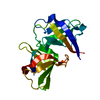
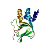
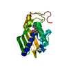
| Structure viewer | Molecule: MolmilJmol/JSmol |
|---|
- Downloads & links
-Download
| PDBx/mmCIF format | 1ryl.cif.gz | 77.1 KB | Display | PDBx/mmCIF format |
|---|---|---|---|---|
| PDB format | pdb1ryl.ent.gz | 58.8 KB | Display | PDB format |
| PDBx/mmJSON format | 1ryl.json.gz | Tree view | PDBx/mmJSON format | |
| Others |  Other downloads Other downloads |
-Validation report
| Arichive directory | https://data.pdbj.org/pub/pdb/validation_reports/ry/1rylftp://data.pdbj.org/pub/pdb/validation_reports/ry/1ryl | HTTPS FTP |
|---|
-Related structure data
| Similar structure data | |
|---|---|
| Other databases |


-Links
 PDBj
PDBj- Assembly
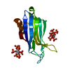
| Deposited unit | 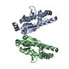
| ||||||||
|---|---|---|---|---|---|---|---|---|---|
| 1 | 
| ||||||||
| 2 | 
| ||||||||
| Unit cell |
| ||||||||
| Details | The biological assembly is a monomer. MolA and MolB represent two molecules in asymmetric unit. |
-Components
| #1: Protein | Mass: 19040.639 Da / Num. of mol.: 2 Source method: isolated from a genetically manipulated source Source: (gene. exp.) #2: Water | ChemComp-HOH / |  Mass: 18.015 Da / Num. of mol.: 259 / Source method: isolated from a natural source / Formula: H2O Mass: 18.015 Da / Num. of mol.: 259 / Source method: isolated from a natural source / Formula: H2O |
|---|
-Experimental details
-Experiment
| Experiment | Method: X-RAY DIFFRACTION / Number of used crystals: 1 |
|---|
- Sample preparation
| Crystal | Density Matthews: 2.17 Å3/Da / Density % sol: 43.32 % |
|---|---|
| Crystal grow | Temperature: 298 K / Method: vapor diffusion, hanging drop / pH: 7 Details: 0.2 M MgCl2, 0.1 M HEPES, 30%PEG3350, 8% Ethanol, pH 7, VAPOR DIFFUSION, HANGING DROP, temperature 298K |
-Data collection
| Diffraction | Mean temperature: 100 K | ||||||||||||
|---|---|---|---|---|---|---|---|---|---|---|---|---|---|
| Diffraction source | Source: SYNCHROTRON / Site: APS  / Beamline: 19-ID / Wavelength: 0.9795,0.9797,0.94656 / Beamline: 19-ID / Wavelength: 0.9795,0.9797,0.94656 | ||||||||||||
| Detector | Type: SBC-2 / Detector: CCD / Date: Dec 12, 2003 / Details: mirrors | ||||||||||||
| Radiation | Monochromator: Si 111 CHANNEL / Protocol: MAD / Monochromatic (M) / Laue (L): M / Scattering type: x-ray | ||||||||||||
| Radiation wavelength |
| ||||||||||||
| Reflection | Resolution: 1.6→50 Å / Num. all: 84444 / Num. obs: 78702 / % possible obs: 93.2 % / Observed criterion σ(F): 2 / Observed criterion σ(I): 4 / Redundancy: 2.37 % / Biso Wilson estimate: 17.2 Å2 / Rmerge(I) obs: 0.055 / Net I/σ(I): 15.85 | ||||||||||||
| Reflection shell | Resolution: 1.6→1.66 Å / Redundancy: 1.3 % / Rmerge(I) obs: 0.372 / Mean I/σ(I) obs: 1.223 / Num. unique all: 4690 / % possible all: 55.5 |
- Processing
| Software |
| |||||||||||||||||||||||||
|---|---|---|---|---|---|---|---|---|---|---|---|---|---|---|---|---|---|---|---|---|---|---|---|---|---|---|
| Refinement | Method to determine structure: MAD / Resolution: 1.6→30.23 Å / Rfactor Rfree error: 0.004 / Data cutoff high absF: 441847.86 / Data cutoff low absF: 0 / Isotropic thermal model: RESTRAINED / Cross valid method: THROUGHOUT / σ(F): 0 / Stereochemistry target values: Engh & Huber
| |||||||||||||||||||||||||
| Solvent computation | Solvent model: FLAT MODEL / Bsol: 57.6538 Å2 / ksol: 0.410761 e/Å3 | |||||||||||||||||||||||||
| Displacement parameters | Biso mean: 22.3 Å2
| |||||||||||||||||||||||||
| Refine analyze |
| |||||||||||||||||||||||||
| Refinement step | Cycle: LAST / Resolution: 1.6→30.23 Å
| |||||||||||||||||||||||||
| Refine LS restraints |
| |||||||||||||||||||||||||
| LS refinement shell | Resolution: 1.6→1.7 Å / Rfactor Rfree error: 0.019 / Total num. of bins used: 6
| |||||||||||||||||||||||||
| Xplor file |
|
