| 登録情報 | データベース: PDB / ID: 6lcy
|
|---|
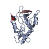
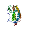
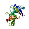
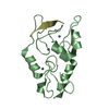
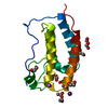
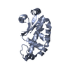
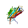
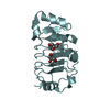
| タイトル | Crystal structure of Synaptotagmin-7 C2B in complex with IP6 |
|---|
 要素 要素 | Synaptotagmin-7 |
|---|
 キーワード キーワード | EXOCYTOSIS / Synaptotagmin-7 / IP6 / Insulin secretion |
|---|
| 機能・相同性 |  機能・相同性情報 機能・相同性情報
calcium ion regulated lysosome exocytosis / vesicle-mediated cholesterol transport / regulation of glucagon secretion / regulation of bone remodeling / phagosome-lysosome fusion / calcium-dependent activation of synaptic vesicle fusion / calcium ion-regulated exocytosis of neurotransmitter / regulation of calcium ion-dependent exocytosis / synaptic vesicle recycling / calcium ion sensor activity ...calcium ion regulated lysosome exocytosis / vesicle-mediated cholesterol transport / regulation of glucagon secretion / regulation of bone remodeling / phagosome-lysosome fusion / calcium-dependent activation of synaptic vesicle fusion / calcium ion-regulated exocytosis of neurotransmitter / regulation of calcium ion-dependent exocytosis / synaptic vesicle recycling / calcium ion sensor activity / short-term synaptic potentiation / positive regulation of calcium ion-dependent exocytosis / calcium-ion regulated exocytosis / vesicle fusion / calcium-dependent phospholipid binding / dense core granule / regulation of phagocytosis / early phagosome / plasma membrane repair / syntaxin binding / peroxisomal membrane / insulin secretion / phosphatidylserine binding / clathrin binding / regulation of insulin secretion / regulation of dopamine secretion / regulation of synaptic vesicle endocytosis / phagocytosis / vesicle-mediated transport / phosphatidylinositol-4,5-bisphosphate binding / axon terminus / hippocampal mossy fiber to CA3 synapse / SNARE binding / GABA-ergic synapse / phagocytic vesicle membrane / synaptic vesicle / peroxisome / synaptic vesicle membrane / presynaptic membrane / calmodulin binding / lysosome / axon / lysosomal membrane / neuronal cell body / calcium ion binding / synapse / dendrite / glutamatergic synapse / plasma membrane / cytosol類似検索 - 分子機能 Synaptotagmin 7, C2A domain / Synaptotagmin 7, C2B domain / Synaptotagmin / Protein kinase C conserved region 2 (CalB) / C2 domain / C2 domain / C2 domain profile. / C2 domain superfamily類似検索 - ドメイン・相同性 |
|---|
| 生物種 |   Mus musculus (ハツカネズミ) Mus musculus (ハツカネズミ) |
|---|
| 手法 |  X線回折 / シンクロトロン / 分子置換 / 解像度: 2.301 Å X線回折 / シンクロトロン / 分子置換 / 解像度: 2.301 Å |
|---|
 データ登録者 データ登録者 | Zhang, Y. / Zhang, X. / Rao, F. / Wang, C. |
|---|
 引用 引用 | ジャーナル: Nat Metab / 年: 2021
タイトル: 5-IP 7 is a GPCR messenger mediating neural control of synaptotagmin-dependent insulin exocytosis and glucose homeostasis.
著者: Zhang, X. / Li, N. / Zhang, J. / Zhang, Y. / Yang, X. / Luo, Y. / Zhang, B. / Xu, Z. / Zhu, Z. / Yang, X. / Yan, Y. / Lin, B. / Wang, S. / Chen, D. / Ye, C. / Ding, Y. / Lou, M. / Wu, Q. / ...著者: Zhang, X. / Li, N. / Zhang, J. / Zhang, Y. / Yang, X. / Luo, Y. / Zhang, B. / Xu, Z. / Zhu, Z. / Yang, X. / Yan, Y. / Lin, B. / Wang, S. / Chen, D. / Ye, C. / Ding, Y. / Lou, M. / Wu, Q. / Hou, Z. / Zhang, K. / Liang, Z. / Wei, A. / Wang, B. / Wang, C. / Jiang, N. / Zhang, W. / Xiao, G. / Ma, C. / Ren, Y. / Qi, X. / Han, W. / Wang, C. / Rao, F. |
|---|
| 履歴 | | 登録 | 2019年11月20日 | 登録サイト: PDBJ / 処理サイト: PDBJ |
|---|
| 改定 1.0 | 2021年3月3日 | Provider: repository / タイプ: Initial release |
|---|
| 改定 1.1 | 2021年11月17日 | Group: Database references / カテゴリ: citation / citation_author / database_2
Item: _citation.journal_abbrev / _citation.journal_id_CSD ..._citation.journal_abbrev / _citation.journal_id_CSD / _citation.journal_id_ISSN / _citation.journal_volume / _citation.page_first / _citation.page_last / _citation.pdbx_database_id_DOI / _citation.pdbx_database_id_PubMed / _citation.title / _citation.year / _database_2.pdbx_DOI / _database_2.pdbx_database_accession |
|---|
| 改定 1.2 | 2023年11月22日 | Group: Data collection / Refinement description
カテゴリ: chem_comp_atom / chem_comp_bond / pdbx_initial_refinement_model |
|---|
|
|---|
 ムービー
ムービー コントローラー


 データを開く
 基本情報
基本情報 構造の表示
構造の表示 ダウンロードとリンク
 その他のダウンロード
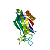


 PDBj
PDBj


 集合体
集合体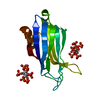


 分子量: 660.035 Da / 分子数: 2 / 由来タイプ: 合成 / 式: C6H18O24P6 / タイプ: SUBJECT OF INVESTIGATION
 分子量: 18.015 Da / 分子数: 34 / 由来タイプ: 天然 / 式: H2O
 試料調製
試料調製 / ビームライン: BL17U1 / 波長: 0.97918 Å
 解析
解析