 Movie
Movie Controller


+ Open data

- Basic information
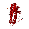
| Entry | Database: PDB / ID: 4eve | ||||||
|---|---|---|---|---|---|---|---|
| Title | Crystal Structure HP-NAP from strain YS29 in apo form | ||||||
 Components Components | Neutrophil-activating protein | ||||||
 Keywords Keywords | METAL TRANSPORT / dodecamer / four-helix bundle | ||||||
| Function / homology |  Function and homology information Function and homology informationoxidoreductase activity, acting on metal ions / ferric iron binding / cytoplasm Similarity search - Function | ||||||
| Biological species |   Helicobacter pylori (bacteria) Helicobacter pylori (bacteria) | ||||||
| Method |  X-RAY DIFFRACTION / SYNCHROTRON / MOLECULAR REPLACEMENT / Resolution: 2.1 Å X-RAY DIFFRACTION / SYNCHROTRON / MOLECULAR REPLACEMENT / Resolution: 2.1 Å | ||||||
 Authors Authors | Yokoyama, H. / Tsuruta, O. / Akao, N. / Fujii, S. | ||||||
 Citation Citation | Journal: Biochem.Biophys.Res.Commun. / Year: 2012 Title: Crystal structure of Helicobacter pylori neutrophil-activating protein with a di-nuclear ferroxidase center in a zinc or cadmium-bound form Authors: Yokoyama, H. / Tsuruta, O. / Akao, N. / Fujii, S. #1: Journal: Acta Crystallogr.,Sect.F / Year: 2012Title: A new crystal lattice structure of Helicobacter pylori neutrophil-activating protein (HP-NAP) Authors: Tsuruta, O. / Yokoyama, H. / Fujii, S. | ||||||
| History |
|
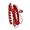
- Structure visualization
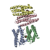
| Structure viewer | Molecule: MolmilJmol/JSmol |
|---|
- Downloads & links
-Download
| PDBx/mmCIF format | 4eve.cif.gz | 46 KB | Display | PDBx/mmCIF format |
|---|---|---|---|---|
| PDB format | pdb4eve.ent.gz | 33.4 KB | Display | PDB format |
| PDBx/mmJSON format | 4eve.json.gz | Tree view | PDBx/mmJSON format | |
| Others |  Other downloads Other downloads |
-Validation report
| Arichive directory | https://data.pdbj.org/pub/pdb/validation_reports/ev/4eveftp://data.pdbj.org/pub/pdb/validation_reports/ev/4eve | HTTPS FTP |
|---|
-Related structure data
| Related structure data | 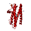 4evbC  4evcC 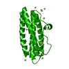 4evdC  3ta8S S: Starting model for refinement C: citing same article ( |
|---|---|
| Similar structure data |


-Links
 PDBj
PDBj


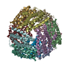
- Assembly
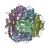
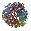
| Deposited unit | 
| ||||||||||||
|---|---|---|---|---|---|---|---|---|---|---|---|---|---|
| 1 | x 12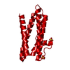
| ||||||||||||
| Unit cell |
| ||||||||||||
| Components on special symmetry positions |
|
-Components
| #1: Protein | Mass: 19106.736 Da / Num. of mol.: 1 Source method: isolated from a genetically manipulated source Source: (gene. exp.) Helicobacter pylori (bacteria) / Strain: YS29 / Gene: napA / Plasmid: pET15b / Production host: | ||
|---|---|---|---|
| #2: Chemical |   Mass: 96.063 Da / Num. of mol.: 2 / Source method: obtained synthetically / Formula: SO4 Mass: 96.063 Da / Num. of mol.: 2 / Source method: obtained synthetically / Formula: SO4#3: Water | ChemComp-HOH / |  Mass: 18.015 Da / Num. of mol.: 122 / Source method: isolated from a natural source / Formula: H2O Mass: 18.015 Da / Num. of mol.: 122 / Source method: isolated from a natural source / Formula: H2O |
-Experimental details
-Experiment
| Experiment | Method: X-RAY DIFFRACTION / Number of used crystals: 1 |
|---|
- Sample preparation
| Crystal | Density Matthews: 3.61 Å3/Da / Density % sol: 65.93 % |
|---|---|
| Crystal grow | Temperature: 293 K / Method: vapor diffusion, sitting drop / pH: 7.5 Details: 2.0M ammonium sulfate, 0.1M Tris-HCl, 0.1M L-Arginine, pH 7.5, VAPOR DIFFUSION, SITTING DROP, temperature 293K |
-Data collection
| Diffraction | Mean temperature: 95 K |
|---|---|
| Diffraction source | Source: SYNCHROTRON / Site: Photon Factory  / Beamline: BL-6A / Wavelength: 0.978 Å / Beamline: BL-6A / Wavelength: 0.978 Å |
| Detector | Type: ADSC QUANTUM 4r / Detector: CCD / Date: Nov 8, 2007 / Details: mirrors |
| Radiation | Monochromator: SI(111) / Protocol: SINGLE WAVELENGTH / Monochromatic (M) / Laue (L): M / Scattering type: x-ray |
| Radiation wavelength | Wavelength: 0.978 Å / Relative weight: 1 |
| Reflection | Resolution: 2.1→20 Å / Num. obs: 16933 / % possible obs: 98.7 % / Redundancy: 9.7 % / Biso Wilson estimate: 25.1 Å2 / Rmerge(I) obs: 0.054 / Net I/σ(I): 44.1 |
| Reflection shell | Resolution: 2.1→2.17 Å / Rmerge(I) obs: 0.301 / Mean I/σ(I) obs: 9.2 / % possible all: 97.4 |
- Processing
| Software |
| |||||||||||||||||||||||||||||||||||||||||||||||||||||||||||||||||
|---|---|---|---|---|---|---|---|---|---|---|---|---|---|---|---|---|---|---|---|---|---|---|---|---|---|---|---|---|---|---|---|---|---|---|---|---|---|---|---|---|---|---|---|---|---|---|---|---|---|---|---|---|---|---|---|---|---|---|---|---|---|---|---|---|---|---|
| Refinement | Method to determine structure: MOLECULAR REPLACEMENT Starting model: PDB ENTRY 3TA8 Resolution: 2.1→19.69 Å / Cor.coef. Fo:Fc: 0.935 / Cor.coef. Fo:Fc free: 0.913 / Occupancy max: 1 / Occupancy min: 0.25 / SU B: 3.567 / SU ML: 0.096 / Cross valid method: THROUGHOUT / σ(F): 0 / ESU R: 0.174 / ESU R Free: 0.163 / Stereochemistry target values: MAXIMUM LIKELIHOOD Details: HYDROGENS HAVE BEEN ADDED IN THE RIDING POSITIONS U VALUES: REFINED INDIVIDUALLY
| |||||||||||||||||||||||||||||||||||||||||||||||||||||||||||||||||
| Solvent computation | Ion probe radii: 0.8 Å / Shrinkage radii: 0.8 Å / VDW probe radii: 1.4 Å / Solvent model: MASK | |||||||||||||||||||||||||||||||||||||||||||||||||||||||||||||||||
| Displacement parameters | Biso max: 53.98 Å2 / Biso mean: 23.1644 Å2 / Biso min: 13.12 Å2 | |||||||||||||||||||||||||||||||||||||||||||||||||||||||||||||||||
| Refine analyze |
| |||||||||||||||||||||||||||||||||||||||||||||||||||||||||||||||||
| Refinement step | Cycle: LAST / Resolution: 2.1→19.69 Å
| |||||||||||||||||||||||||||||||||||||||||||||||||||||||||||||||||
| Refine LS restraints |
| |||||||||||||||||||||||||||||||||||||||||||||||||||||||||||||||||
| LS refinement shell | Resolution: 2.099→2.153 Å / Total num. of bins used: 20
|
