 Movie
Movie Controller

[English] 日本語
 Yorodumi
- PDB-3hqh: Structures of SPOP-Substrate Complexes: Insights into Molecular A... -
+ Open data

- Basic information
| Entry | Database: PDB / ID: 3hqh | ||||||
|---|---|---|---|---|---|---|---|
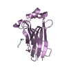
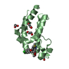
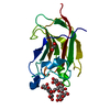
| Title | Structures of SPOP-Substrate Complexes: Insights into Molecular Architectures of BTB-Cul3 Ubiquitin Ligases: SPOPMATHx-MacroH2ASBCpep1 | ||||||
 Components Components |
| ||||||
 Keywords Keywords | LIGASE / ubiquitin / SPOP / BTB / E3 / Nucleus / Ubl conjugation pathway | ||||||
| Function / homology |  Function and homology information Function and homology informationHedgehog 'on' state / negative regulation of cell cycle G2/M phase transition / positive regulation of endodermal cell differentiation / negative regulation of protein localization to chromosome, telomeric region / regulation of NAD metabolic process / positive regulation of response to oxidative stress / Barr body / positive regulation of maintenance of mitotic sister chromatid cohesion / regulation of response to oxidative stress / ADP-D-ribose binding ...Hedgehog 'on' state / negative regulation of cell cycle G2/M phase transition / positive regulation of endodermal cell differentiation / negative regulation of protein localization to chromosome, telomeric region / regulation of NAD metabolic process / positive regulation of response to oxidative stress / Barr body / positive regulation of maintenance of mitotic sister chromatid cohesion / regulation of response to oxidative stress / ADP-D-ribose binding / ADP-D-ribose modification-dependent protein binding / negative regulation of transcription of nucleolar large rRNA by RNA polymerase I / double-stranded methylated DNA binding / regulation of oxidative phosphorylation / sex chromatin / rDNA binding / positive regulation of keratinocyte differentiation / establishment of protein localization to chromatin / dosage compensation by inactivation of X chromosome / poly-ADP-D-ribose modification-dependent protein binding / negative regulation of response to oxidative stress / molecular function inhibitor activity / protein poly-ADP-ribosylation / nucleosomal DNA binding / Cul3-RING ubiquitin ligase complex / nuclear chromosome / negative regulation of gene expression, epigenetic / regulation of lipid metabolic process / regulation of proteolysis / protein serine/threonine kinase inhibitor activity / pericentric heterochromatin / site of DNA damage / condensed chromosome / epigenetic regulation of gene expression / transcription initiation-coupled chromatin remodeling / RNA polymerase II transcription regulatory region sequence-specific DNA binding / promoter-specific chromatin binding / chromatin DNA binding / Hedgehog 'on' state / protein polyubiquitination / structural constituent of chromatin / nucleosome / nucleosome assembly / heterochromatin formation / proteasome-mediated ubiquitin-dependent protein catabolic process / chromosome, telomeric region / nuclear speck / transcription cis-regulatory region binding / protein ubiquitination / protein heterodimerization activity / DNA repair / ubiquitin protein ligase binding / protein kinase binding / nucleolus / chromatin / enzyme binding / negative regulation of transcription by RNA polymerase II / DNA binding / extracellular exosome / nucleoplasm / identical protein binding / nucleus / cytoplasm Similarity search - Function | ||||||
| Biological species |  Homo sapiens (human) Homo sapiens (human) | ||||||
| Method |  X-RAY DIFFRACTION / SYNCHROTRON / MOLECULAR REPLACEMENT / Resolution: 2.3 Å X-RAY DIFFRACTION / SYNCHROTRON / MOLECULAR REPLACEMENT / Resolution: 2.3 Å | ||||||
 Authors Authors | Zhuang, M. / Schulman, B.A. | ||||||
 Citation Citation | Journal: Mol.Cell / Year: 2009 Title: Structures of SPOP-substrate complexes: insights into molecular architectures of BTB-Cul3 ubiquitin ligases. Authors: Zhuang, M. / Calabrese, M.F. / Liu, J. / Waddell, M.B. / Nourse, A. / Hammel, M. / Miller, D.J. / Walden, H. / Duda, D.M. / Seyedin, S.N. / Hoggard, T. / Harper, J.W. / White, K.P. / Schulman, B.A. | ||||||
| History |
|
- Structure visualization
| Structure viewer | Molecule: MolmilJmol/JSmol |
|---|
- Downloads & links
-Download
| PDBx/mmCIF format | 3hqh.cif.gz | 43.5 KB | Display | PDBx/mmCIF format |
|---|---|---|---|---|
| PDB format | pdb3hqh.ent.gz | 29.4 KB | Display | PDB format |
| PDBx/mmJSON format | 3hqh.json.gz | Tree view | PDBx/mmJSON format | |
| Others |  Other downloads Other downloads |
-Validation report
| Arichive directory | https://data.pdbj.org/pub/pdb/validation_reports/hq/3hqhftp://data.pdbj.org/pub/pdb/validation_reports/hq/3hqh | HTTPS FTP |
|---|
-Related structure data
| Related structure data | 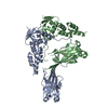 3hqiC 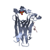 3hqlC  3hqmC  3hsvC  3htmC  3hu6C 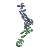 3hveC  3ivqC 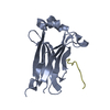 3ivvC  2cr2S C: citing same article ( S: Starting model for refinement |
|---|---|
| Similar structure data |


-Links
 PDBj
PDBj


- Assembly
| Deposited unit | 
| ||||||||
|---|---|---|---|---|---|---|---|---|---|
| 1 |
| ||||||||
| Unit cell |
|
-Components
| #1: Protein | Mass: 16485.932 Da / Num. of mol.: 1 Source method: isolated from a genetically manipulated source Source: (gene. exp.) Homo sapiens (human) / Gene: spop / Production host:  | ||
|---|---|---|---|
| #2: Protein/peptide | Mass: 1422.429 Da / Num. of mol.: 1 / Source method: obtained synthetically / Details: synthetic peptide / References: UniProt: O75367*PLUS | ||
| #3: Chemical |   Mass: 65.409 Da / Num. of mol.: 3 / Source method: obtained synthetically / Formula: Zn Mass: 65.409 Da / Num. of mol.: 3 / Source method: obtained synthetically / Formula: Zn#4: Water | ChemComp-HOH / |  Mass: 18.015 Da / Num. of mol.: 48 / Source method: isolated from a natural source / Formula: H2O Mass: 18.015 Da / Num. of mol.: 48 / Source method: isolated from a natural source / Formula: H2O |
-Experimental details
-Experiment
| Experiment | Method: X-RAY DIFFRACTION / Number of used crystals: 1 |
|---|
- Sample preparation
| Crystal | Density Matthews: 2.19 Å3/Da / Density % sol: 43.82 % |
|---|---|
| Crystal grow | Temperature: 291 K / Method: vapor diffusion, hanging drop / pH: 6.5 Details: 12% PEG550, 0.1M MES 6.5, 20mM ZnSO4 , VAPOR DIFFUSION, HANGING DROP, temperature 291K |
-Data collection
| Diffraction source | Source: SYNCHROTRON / Site: APS  / Beamline: 22-BM / Wavelength: 1 Å / Beamline: 22-BM / Wavelength: 1 Å |
|---|---|
| Detector | Type: MARMOSAIC 225 mm CCD / Detector: CCD |
| Radiation | Protocol: SINGLE WAVELENGTH / Monochromatic (M) / Laue (L): M / Scattering type: x-ray |
| Radiation wavelength | Wavelength: 1 Å / Relative weight: 1 |
| Reflection | Resolution: 2.29→50 Å / Num. all: 8052 / Num. obs: 8052 / Observed criterion σ(F): 0 / Observed criterion σ(I): 8.3 / Redundancy: 7 % / Rmerge(I) obs: 0.09 / Rsym value: 0.09 / Net I/σ(I): 31.7 |
| Reflection shell | Resolution: 2.29→2.37 Å / Redundancy: 4.5 % / Mean I/σ(I) obs: 8.3 / Rsym value: 0.182 |
- Processing
| Software |
| |||||||||||||||||||||||||
|---|---|---|---|---|---|---|---|---|---|---|---|---|---|---|---|---|---|---|---|---|---|---|---|---|---|---|
| Refinement | Method to determine structure: MOLECULAR REPLACEMENT Starting model: PDB entry 2cr2.pdb Resolution: 2.3→50 Å / Cross valid method: Rfree / σ(F): 0 / σ(I): 8.3
| |||||||||||||||||||||||||
| Refinement step | Cycle: LAST / Resolution: 2.3→50 Å
|
