- PDB-6f8f: Co-crystal structure of SPOP MATH domain and human Pdx1 fragment -
+
Open data
ID or keywords:
Loading...
-
Basic information
Entry
Database: PDB / ID: 6f8f
Title
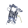
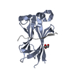
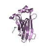
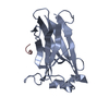
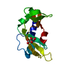
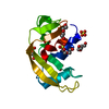
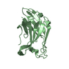
Co-crystal structure of SPOP MATH domain and human Pdx1 fragment
Components
Pancreas/duodenum homeobox protein 1
Speckle-type POZ protein
Keywords
LIGASE / Nuclear / Diabetes
Function / homology
Function and homology information
response to vitamin / type B pancreatic cell differentiation / negative regulation of type B pancreatic cell apoptotic process / transdifferentiation / Regulation of gene expression in early pancreatic precursor cells / response to chlorate / positive regulation of type B pancreatic cell proliferation / glucose mediated signaling pathway / type B pancreatic cell apoptotic process / morphogenesis of embryonic epithelium ...response to vitamin / type B pancreatic cell differentiation / negative regulation of type B pancreatic cell apoptotic process / transdifferentiation / Regulation of gene expression in early pancreatic precursor cells / response to chlorate / positive regulation of type B pancreatic cell proliferation / glucose mediated signaling pathway / type B pancreatic cell apoptotic process / morphogenesis of embryonic epithelium / response to fatty acid / response to L-leucine / response to alkaloid / exocrine pancreas development / type B pancreatic cell proliferation / digestive tract development / insulin secretion / molecular function inhibitor activity / smoothened signaling pathway / Cul3-RING ubiquitin ligase complex / response to iron(II) ion / Regulation of gene expression in beta cells / regulation of proteolysis / intrinsic apoptotic signaling pathway in response to endoplasmic reticulum stress / animal organ regeneration / negative regulation of endoplasmic reticulum stress-induced intrinsic apoptotic signaling pathway / response to cytokine / response to glucocorticoid / animal organ morphogenesis / positive regulation of insulin secretion involved in cellular response to glucose stimulus / stem cell differentiation / response to nicotine / generation of precursor metabolites and energy / liver development / promoter-specific chromatin binding / Hedgehog 'on' state / glucose metabolic process / negative regulation of epithelial cell proliferation / positive regulation of insulin secretion / sequence-specific double-stranded DNA binding / protein polyubiquitination / transcription by RNA polymerase II / proteasome-mediated ubiquitin-dependent protein catabolic process / DNA-binding transcription factor activity, RNA polymerase II-specific / nuclear speck / RNA polymerase II cis-regulatory region sequence-specific DNA binding / DNA-binding transcription factor activity / response to xenobiotic stimulus / ubiquitin protein ligase binding / regulation of transcription by RNA polymerase II / chromatin / negative regulation of transcription by RNA polymerase II / positive regulation of transcription by RNA polymerase II / nucleoplasm / identical protein binding / nucleus / cytoplasm / cytosol Similarity search - Function
Homeobox protein, antennapedia type / MATH domain / SPOP, C-terminal BACK domain / : / BPM/SPOP, BACK domain / Apoptosis, Tumor Necrosis Factor Receptor Associated Protein 2; Chain A / Apoptosis, Tumor Necrosis Factor Receptor Associated Protein 2; Chain A / Homeobox domain, metazoa / MATH domain / MATH/TRAF domain ...Homeobox protein, antennapedia type / MATH domain / SPOP, C-terminal BACK domain / : / BPM/SPOP, BACK domain / Apoptosis, Tumor Necrosis Factor Receptor Associated Protein 2; Chain A / Apoptosis, Tumor Necrosis Factor Receptor Associated Protein 2; Chain A / Homeobox domain, metazoa / MATH domain / MATH/TRAF domain / MATH/TRAF domain profile. / meprin and TRAF homology / TRAF-like / Homeobox, conserved site / 'Homeobox' domain signature. / Homeodomain / 'Homeobox' domain profile. / Homeodomain / Homeobox domain / BTB/POZ domain / BTB domain profile. / Broad-Complex, Tramtrack and Bric a brac / BTB/POZ domain / SKP1/BTB/POZ domain superfamily / Homeobox-like domain superfamily / Sandwich / Mainly Beta Similarity search - Domain/homology
Resolution: 2→19.94 Å / Cor.coef. Fo:Fc: 0.969 / Cor.coef. Fo:Fc free: 0.945 / SU B: 7.211 / SU ML: 0.183 / Cross valid method: THROUGHOUT / ESU R: 0.176 / ESU R Free: 0.175 / Stereochemistry target values: MAXIMUM LIKELIHOOD / Details: HYDROGENS HAVE BEEN ADDED IN THE RIDING POSITIONS
Rfactor
Num. reflection
% reflection
Selection details
Rfree
0.27433
640
5 %
RANDOM
Rwork
0.21549
-
-
-
obs
0.21841
12184
99.07 %
-
Solvent computation
Ion probe radii: 0.8 Å / Shrinkage radii: 0.8 Å / VDW probe radii: 1.2 Å / Solvent model: MASK
 Movie
Movie Controller


 Open data
 Basic information
 Components
 Keywords
 Function and homology information
 Homo sapiens (human)
 X-RAY DIFFRACTION /
  Authors
 Citation
 Structure visualization
 Downloads & links
 Other downloads


 PDBj
PDBj


 Assembly

 Mass: 18.015 Da / Num. of mol.: 35 / Source method: isolated from a natural source / Formula: H2O
 Sample preparation
 / Beamline: ID30B / Wavelength: 0.82655 Å
 Processing