 Movie
Movie Controller

[English] 日本語
 Yorodumi
- PDB-5f3y: Crystal Structure of Myo7b N-MyTH4-FERM-SH3 in complex with Anks4b CEN -
+ Open data

- Basic information
| Entry | Database: PDB / ID: 5f3y | ||||||
|---|---|---|---|---|---|---|---|
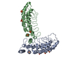
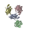
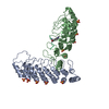
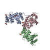
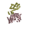
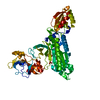
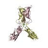
| Title | Crystal Structure of Myo7b N-MyTH4-FERM-SH3 in complex with Anks4b CEN | ||||||
 Components Components |
| ||||||
 Keywords Keywords | MOTOR PROTEIN/PROTEIN BINDING / MOTOR PROTEIN / PROTEIN BINDING / STRUCTURAL PROTEIN / PROTEIN TRANSPORT / MOTOR PROTEIN-PROTEIN BINDING complex | ||||||
| Function / homology |  Function and homology information Function and homology informationprotein localization to microvillus / apical cytoplasm / brush border assembly / stereocilia ankle link / myosin complex / myosin binding / microvillus / cytoskeletal motor activity / brush border / response to endoplasmic reticulum stress ...protein localization to microvillus / apical cytoplasm / brush border assembly / stereocilia ankle link / myosin complex / myosin binding / microvillus / cytoskeletal motor activity / brush border / response to endoplasmic reticulum stress / actin binding / protein-containing complex assembly / cell differentiation / endoplasmic reticulum membrane / ATP binding / plasma membrane Similarity search - Function | ||||||
| Biological species |  | ||||||
| Method |  X-RAY DIFFRACTION / SYNCHROTRON / MOLECULAR REPLACEMENT / Resolution: 3.409 Å X-RAY DIFFRACTION / SYNCHROTRON / MOLECULAR REPLACEMENT / Resolution: 3.409 Å | ||||||
 Authors Authors | Li, J. / He, Y. / Lu, Q. / Zhang, M. | ||||||
 Citation Citation | Journal: Dev.Cell / Year: 2016 Title: Mechanistic Basis of Organization of the Harmonin/USH1C-Mediated Brush Border Microvilli Tip-Link Complex Authors: Li, J. / He, Y. / Lu, Q. / Zhang, M. | ||||||
| History |
|
- Structure visualization
| Structure viewer | Molecule: MolmilJmol/JSmol |
|---|
- Downloads & links
-Download
| PDBx/mmCIF format | 5f3y.cif.gz | 227.8 KB | Display | PDBx/mmCIF format |
|---|---|---|---|---|
| PDB format | pdb5f3y.ent.gz | 178.3 KB | Display | PDB format |
| PDBx/mmJSON format | 5f3y.json.gz | Tree view | PDBx/mmJSON format | |
| Others |  Other downloads Other downloads |
-Validation report
| Arichive directory | https://data.pdbj.org/pub/pdb/validation_reports/f3/5f3yftp://data.pdbj.org/pub/pdb/validation_reports/f3/5f3y | HTTPS FTP |
|---|
-Related structure data
| Related structure data |  5f3xC 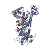 3pvlS S: Starting model for refinement C: citing same article ( |
|---|---|
| Similar structure data |

-Links
 PDBj
PDBj


- Assembly
| Deposited unit | 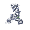
| ||||||||
|---|---|---|---|---|---|---|---|---|---|
| 1 |
| ||||||||
| Unit cell |
|
-Components
| #1: Protein | Mass: 69985.125 Da / Num. of mol.: 1 / Fragment: N-MyTH4-FERM-SH3, UNP residues 962-1578 Source method: isolated from a genetically manipulated source Source: (gene. exp.)  |
|---|---|
| #2: Protein | Mass: 11613.118 Da / Num. of mol.: 1 / Fragment: CEN, UNP residues 252-352 Source method: isolated from a genetically manipulated source Source: (gene. exp.) |
-Experimental details
-Experiment
| Experiment | Method: X-RAY DIFFRACTION / Number of used crystals: 1 |
|---|
- Sample preparation
| Crystal | Density Matthews: 5.84 Å3/Da / Density % sol: 78.94 % |
|---|---|
| Crystal grow | Temperature: 289 K / Method: vapor diffusion, hanging drop / pH: 6.5 Details: 25%(w/v) Pentaerythritol ethoxylate (3/4 EO/OH), 0.1 M MES PH range: 6.5 |
-Data collection
| Diffraction | Mean temperature: 100 K | |||||||||||||||||||||||||||||||||||||||||||||||||||||||||||||||||||||||||||||||||||||||||||||||||||||||||||||||||||||||||||||||||||||||||||||||||||||||||||||||||||||||||||||||||||||||||||||
|---|---|---|---|---|---|---|---|---|---|---|---|---|---|---|---|---|---|---|---|---|---|---|---|---|---|---|---|---|---|---|---|---|---|---|---|---|---|---|---|---|---|---|---|---|---|---|---|---|---|---|---|---|---|---|---|---|---|---|---|---|---|---|---|---|---|---|---|---|---|---|---|---|---|---|---|---|---|---|---|---|---|---|---|---|---|---|---|---|---|---|---|---|---|---|---|---|---|---|---|---|---|---|---|---|---|---|---|---|---|---|---|---|---|---|---|---|---|---|---|---|---|---|---|---|---|---|---|---|---|---|---|---|---|---|---|---|---|---|---|---|---|---|---|---|---|---|---|---|---|---|---|---|---|---|---|---|---|---|---|---|---|---|---|---|---|---|---|---|---|---|---|---|---|---|---|---|---|---|---|---|---|---|---|---|---|---|---|---|---|---|
| Diffraction source | Source: SYNCHROTRON / Site: SSRF  / Beamline: BL17U / Wavelength: 0.9788 Å / Beamline: BL17U / Wavelength: 0.9788 Å | |||||||||||||||||||||||||||||||||||||||||||||||||||||||||||||||||||||||||||||||||||||||||||||||||||||||||||||||||||||||||||||||||||||||||||||||||||||||||||||||||||||||||||||||||||||||||||||
| Detector | Type: ADSC QUANTUM 315r / Detector: CCD / Date: Sep 14, 2015 | |||||||||||||||||||||||||||||||||||||||||||||||||||||||||||||||||||||||||||||||||||||||||||||||||||||||||||||||||||||||||||||||||||||||||||||||||||||||||||||||||||||||||||||||||||||||||||||
| Radiation | Monochromator: Double crystal / Protocol: SINGLE WAVELENGTH / Monochromatic (M) / Laue (L): M / Scattering type: x-ray | |||||||||||||||||||||||||||||||||||||||||||||||||||||||||||||||||||||||||||||||||||||||||||||||||||||||||||||||||||||||||||||||||||||||||||||||||||||||||||||||||||||||||||||||||||||||||||||
| Radiation wavelength | Wavelength: 0.9788 Å / Relative weight: 1 | |||||||||||||||||||||||||||||||||||||||||||||||||||||||||||||||||||||||||||||||||||||||||||||||||||||||||||||||||||||||||||||||||||||||||||||||||||||||||||||||||||||||||||||||||||||||||||||
| Reflection | Resolution: 3.4→50 Å / Num. obs: 24972 / % possible obs: 92.9 % / Redundancy: 6.8 % / Biso Wilson estimate: 92.1 Å2 / Rmerge(I) obs: 0.1 / Rpim(I) all: 0.04 / Rrim(I) all: 0.109 / Χ2: 1.651 / Net I/av σ(I): 19.612 / Net I/σ(I): 8.7 / Num. measured all: 169291 | |||||||||||||||||||||||||||||||||||||||||||||||||||||||||||||||||||||||||||||||||||||||||||||||||||||||||||||||||||||||||||||||||||||||||||||||||||||||||||||||||||||||||||||||||||||||||||||
| Reflection shell | Diffraction-ID: 1 / Rejects: _
|
- Processing
| Software |
| ||||||||||||||||||||||||||||||||||||||||||||||||||||||||||||||||||||||||||||||||||||||||||||||||||||
|---|---|---|---|---|---|---|---|---|---|---|---|---|---|---|---|---|---|---|---|---|---|---|---|---|---|---|---|---|---|---|---|---|---|---|---|---|---|---|---|---|---|---|---|---|---|---|---|---|---|---|---|---|---|---|---|---|---|---|---|---|---|---|---|---|---|---|---|---|---|---|---|---|---|---|---|---|---|---|---|---|---|---|---|---|---|---|---|---|---|---|---|---|---|---|---|---|---|---|---|---|---|
| Refinement | Method to determine structure: MOLECULAR REPLACEMENT Starting model: 3PVL Resolution: 3.409→30.718 Å / SU ML: 0.53 / Cross valid method: FREE R-VALUE / σ(F): 1.33 / Phase error: 30.08 / Stereochemistry target values: ML
| ||||||||||||||||||||||||||||||||||||||||||||||||||||||||||||||||||||||||||||||||||||||||||||||||||||
| Solvent computation | Shrinkage radii: 0.9 Å / VDW probe radii: 1.11 Å / Solvent model: FLAT BULK SOLVENT MODEL | ||||||||||||||||||||||||||||||||||||||||||||||||||||||||||||||||||||||||||||||||||||||||||||||||||||
| Displacement parameters | Biso max: 199.33 Å2 / Biso mean: 93.211 Å2 / Biso min: 38.97 Å2 | ||||||||||||||||||||||||||||||||||||||||||||||||||||||||||||||||||||||||||||||||||||||||||||||||||||
| Refinement step | Cycle: final / Resolution: 3.409→30.718 Å
| ||||||||||||||||||||||||||||||||||||||||||||||||||||||||||||||||||||||||||||||||||||||||||||||||||||
| Refine LS restraints |
| ||||||||||||||||||||||||||||||||||||||||||||||||||||||||||||||||||||||||||||||||||||||||||||||||||||
| LS refinement shell | Refine-ID: X-RAY DIFFRACTION / Total num. of bins used: 9
| ||||||||||||||||||||||||||||||||||||||||||||||||||||||||||||||||||||||||||||||||||||||||||||||||||||
| Refinement TLS params. | Method: refined / Refine-ID: X-RAY DIFFRACTION
| ||||||||||||||||||||||||||||||||||||||||||||||||||||||||||||||||||||||||||||||||||||||||||||||||||||
| Refinement TLS group |
|
