 Movie
Movie Controller

[English] 日本語
 Yorodumi
- PDB-4bpx: Crystal structure of human primase in complex with the primase- b... -
+ Open data

- Basic information
| Entry | Database: PDB / ID: 4bpx | |||||||||
|---|---|---|---|---|---|---|---|---|---|---|
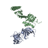
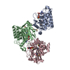
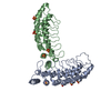
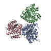
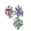
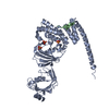
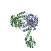
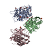
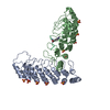
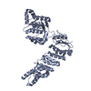
| Title | Crystal structure of human primase in complex with the primase- binding motif of DNA polymerase alpha | |||||||||
 Components Components |
| |||||||||
 Keywords Keywords | TRANSFERASE / DNA REPLICATION / FUSION PROTEIN / CHIMERA | |||||||||
| Function / homology |  Function and homology information Function and homology informationribonucleotide binding / DNA primase AEP / DNA replication initiation / DNA/RNA hybrid binding / Inhibition of replication initiation of damaged DNA by RB1/E2F1 / alpha DNA polymerase:primase complex / regulation of type I interferon production / Telomere C-strand synthesis initiation / Polymerase switching / Processive synthesis on the lagging strand ...ribonucleotide binding / DNA primase AEP / DNA replication initiation / DNA/RNA hybrid binding / Inhibition of replication initiation of damaged DNA by RB1/E2F1 / alpha DNA polymerase:primase complex / regulation of type I interferon production / Telomere C-strand synthesis initiation / Polymerase switching / Processive synthesis on the lagging strand / Removal of the Flap Intermediate / lagging strand elongation / DNA replication, synthesis of primer / mitotic DNA replication initiation / Polymerase switching on the C-strand of the telomere / DNA strand elongation involved in DNA replication / leading strand elongation / G1/S-Specific Transcription / DNA synthesis involved in DNA repair / DNA replication origin binding / Activation of the pre-replicative complex / DNA replication initiation / Defective pyroptosis / double-strand break repair via nonhomologous end joining / nuclear matrix / DNA-directed RNA polymerase activity / nuclear envelope / single-stranded DNA binding / 4 iron, 4 sulfur cluster binding / DNA-directed DNA polymerase / DNA-directed DNA polymerase activity / DNA replication / nucleotide binding / DNA repair / chromatin binding / protein kinase binding / chromatin / nucleolus / magnesium ion binding / DNA binding / zinc ion binding / nucleoplasm / membrane / metal ion binding / nucleus / cytosol Similarity search - Function | |||||||||
| Biological species |  HOMO SAPIENS (human) HOMO SAPIENS (human) | |||||||||
| Method |  X-RAY DIFFRACTION / SYNCHROTRON / MOLECULAR REPLACEMENT / Resolution: 3.4 Å X-RAY DIFFRACTION / SYNCHROTRON / MOLECULAR REPLACEMENT / Resolution: 3.4 Å | |||||||||
 Authors Authors | Kilkenny, M.L. / Perera, R.L. / Pellegrini, L. | |||||||||
 Citation Citation | Journal: Proc.Natl.Acad.Sci.USA / Year: 2013 Title: Structures of Human Primase Reveal Design of Nucleotide Elongation Site and Mode of Pol Alpha Tethering Authors: Kilkenny, M.L. / Longo, M. / Perera, R.L. / Pellegrini, L. | |||||||||
| History |
|
- Structure visualization
| Structure viewer | Molecule: MolmilJmol/JSmol |
|---|
- Downloads & links
-Download
| PDBx/mmCIF format | 4bpx.cif.gz | 459.4 KB | Display | PDBx/mmCIF format |
|---|---|---|---|---|
| PDB format | pdb4bpx.ent.gz | 381.7 KB | Display | PDB format |
| PDBx/mmJSON format | 4bpx.json.gz | Tree view | PDBx/mmJSON format | |
| Others |  Other downloads Other downloads |
-Validation report
| Arichive directory | https://data.pdbj.org/pub/pdb/validation_reports/bp/4bpxftp://data.pdbj.org/pub/pdb/validation_reports/bp/4bpx | HTTPS FTP |
|---|
-Related structure data
| Related structure data |  4bpuSC 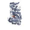 4bpwC S: Starting model for refinement C: citing same article ( |
|---|---|
| Similar structure data |
-Links
 PDBj
PDBj


- Assembly
| Deposited unit | 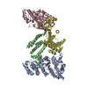
| |||||||||
|---|---|---|---|---|---|---|---|---|---|---|
| 1 | 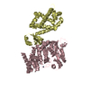
| |||||||||
| 2 | 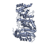
| |||||||||
| 3 | 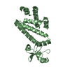
| |||||||||
| Unit cell |
| |||||||||
| Noncrystallographic symmetry (NCS) | NCS domain:
|
-Components
| #1: Protein | Mass: 50108.023 Da / Num. of mol.: 2 / Mutation: YES Source method: isolated from a genetically manipulated source Source: (gene. exp.) HOMO SAPIENS (human) / Production host:  References: UniProt: P49642, Transferases; Transferring phosphorus-containing groups; Nucleotidyltransferases #2: Protein | Mass: 30774.051 Da / Num. of mol.: 2 Fragment: PRIMASE-BINDING MOTIF RESIDUES 1445-1462, PRIL RESIDUES 19-253 Source method: isolated from a genetically manipulated source Details: PRIL IS FUSED AT THE N-TERMINUS TO THE PRIMASE-BINDING MOTIF OF DNA POLYMERASE ALPHA (CHAIN E), VIA A 15-AMINO ACID LINKER OF SEQUENCE TGSTGSTGSTGSTGS Source: (gene. exp.) HOMO SAPIENS (human) / Production host: References: UniProt: P09884, UniProt: P49643, DNA-directed DNA polymerase, Transferases; Transferring phosphorus-containing groups; Nucleotidyltransferases #3: Chemical |   Mass: 65.409 Da / Num. of mol.: 2 / Source method: obtained synthetically / Formula: Zn Mass: 65.409 Da / Num. of mol.: 2 / Source method: obtained synthetically / Formula: ZnSequence details | CHAINS B AND D ARE ENGINEERED, CHIMERIC PROTEIN CONSTRUCTS. IN EACH CONSTRUCT, THE AMINO-TO- ...CHAINS B AND D ARE ENGINEERED | |
|---|
-Experimental details
-Experiment
| Experiment | Method: X-RAY DIFFRACTION / Number of used crystals: 2 |
|---|
- Sample preparation
| Crystal | Density Matthews: 3.9 Å3/Da / Density % sol: 68 % / Description: NONE |
|---|---|
| Crystal grow | Details: 100MM TRIS-HCL/BICINE PH 8.5, 20% GLYCEROL, 10% PEG4000 AND 20MM EACH OF AN ALCOHOL MIX COMPRISING 1,6-HEXANEDIOL, 1-BUTANOL, 1,2-PROPANEDIOL, 2-PROPANOL, 1,4- BUTANEDIOL AND 1,3-PROPANEDIOL. |
-Data collection
| Diffraction | Mean temperature: 100 K |
|---|---|
| Diffraction source | Source: SYNCHROTRON / Site: SOLEIL  / Beamline: PROXIMA 1 / Wavelength: 1.12713 / Beamline: PROXIMA 1 / Wavelength: 1.12713 |
| Detector | Type: DECTRIS PILATUS 6M / Detector: PIXEL / Date: Sep 7, 2012 |
| Radiation | Protocol: SINGLE WAVELENGTH / Monochromatic (M) / Laue (L): M / Scattering type: x-ray |
| Radiation wavelength | Wavelength: 1.12713 Å / Relative weight: 1 |
| Reflection | Resolution: 3.4→49.37 Å / Num. obs: 28805 / % possible obs: 100 % / Observed criterion σ(I): 0 / Redundancy: 9.2 % / Biso Wilson estimate: 170.66 Å2 / Rmerge(I) obs: 0.06 / Net I/σ(I): 13.8 |
| Reflection shell | Resolution: 3.4→3.58 Å / Redundancy: 9.4 % / Rmerge(I) obs: 1.5 / Mean I/σ(I) obs: 1.2 / % possible all: 100 |
- Processing
| Software |
| |||||||||||||||||||||||||||||||||||||||||||||||||||||||||||||||||||||||||||||||||||||||||||||||||||||||||||||||||||||||||||||||||||||||||||||||||||
|---|---|---|---|---|---|---|---|---|---|---|---|---|---|---|---|---|---|---|---|---|---|---|---|---|---|---|---|---|---|---|---|---|---|---|---|---|---|---|---|---|---|---|---|---|---|---|---|---|---|---|---|---|---|---|---|---|---|---|---|---|---|---|---|---|---|---|---|---|---|---|---|---|---|---|---|---|---|---|---|---|---|---|---|---|---|---|---|---|---|---|---|---|---|---|---|---|---|---|---|---|---|---|---|---|---|---|---|---|---|---|---|---|---|---|---|---|---|---|---|---|---|---|---|---|---|---|---|---|---|---|---|---|---|---|---|---|---|---|---|---|---|---|---|---|---|---|---|---|
| Refinement | Method to determine structure: MOLECULAR REPLACEMENT Starting model: PDB ENTRY 4BPU Resolution: 3.4→45.325 Å / SU ML: 0.63 / σ(F): 1.32 / Phase error: 34.39 / Stereochemistry target values: ML Details: THE STRUCTURE WAS REFINED IN PHENIX WITH RIDING HYDROGENS. THE HYDROGENS HAVE BEEN INCLUDED IN THE ENTRY.
| |||||||||||||||||||||||||||||||||||||||||||||||||||||||||||||||||||||||||||||||||||||||||||||||||||||||||||||||||||||||||||||||||||||||||||||||||||
| Solvent computation | Shrinkage radii: 0.9 Å / VDW probe radii: 1.11 Å / Solvent model: FLAT BULK SOLVENT MODEL | |||||||||||||||||||||||||||||||||||||||||||||||||||||||||||||||||||||||||||||||||||||||||||||||||||||||||||||||||||||||||||||||||||||||||||||||||||
| Displacement parameters | Biso mean: 200.1 Å2 | |||||||||||||||||||||||||||||||||||||||||||||||||||||||||||||||||||||||||||||||||||||||||||||||||||||||||||||||||||||||||||||||||||||||||||||||||||
| Refinement step | Cycle: LAST / Resolution: 3.4→45.325 Å
| |||||||||||||||||||||||||||||||||||||||||||||||||||||||||||||||||||||||||||||||||||||||||||||||||||||||||||||||||||||||||||||||||||||||||||||||||||
| Refine LS restraints |
| |||||||||||||||||||||||||||||||||||||||||||||||||||||||||||||||||||||||||||||||||||||||||||||||||||||||||||||||||||||||||||||||||||||||||||||||||||
| LS refinement shell |
|
