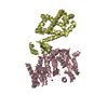
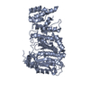
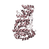
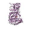
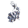
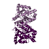
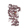
- PDB-4bpw: Crystal structure of human primase bound to UTP -
+
Open data
ID or keywords:
Loading...
-
Basic information
Entry
Database: PDB / ID: 4bpw
Title
Crystal structure of human primase bound to UTP
Components
DNA PRIMASE LARGE SUBUNIT
DNA PRIMASE SMALL SUBUNIT
Keywords
TRANSFERASE / DNA-DEPENDENT RNA POLYMERASE / DNA REPLICATION
Function / homology
Function and homology information
ribonucleotide binding / DNA primase AEP / DNA replication initiation / DNA/RNA hybrid binding / Inhibition of replication initiation of damaged DNA by RB1/E2F1 / Telomere C-strand synthesis initiation / alpha DNA polymerase:primase complex / Polymerase switching / Processive synthesis on the lagging strand / Removal of the Flap Intermediate ...ribonucleotide binding / DNA primase AEP / DNA replication initiation / DNA/RNA hybrid binding / Inhibition of replication initiation of damaged DNA by RB1/E2F1 / Telomere C-strand synthesis initiation / alpha DNA polymerase:primase complex / Polymerase switching / Processive synthesis on the lagging strand / Removal of the Flap Intermediate / DNA replication, synthesis of primer / Polymerase switching on the C-strand of the telomere / Activation of the pre-replicative complex / DNA replication initiation / Defective pyroptosis / DNA-directed RNA polymerase activity / 4 iron, 4 sulfur cluster binding / magnesium ion binding / DNA binding / zinc ion binding / nucleoplasm / membrane / metal ion binding Similarity search - Function
Transcription Elongation Factor S-II; Chain A - #80 / DNA primase, PRIM domain / DNA primase, PRIM domain / : / Eukaryotic and archaeal DNA primase, large subunit N-terminal domain / DNA primase, small subunit, eukaryotic/archaeal / DNA primase large subunit, eukaryotic/archaeal / DNA primase, large subunit, eukaryotic / DNA primase, small subunit / DNA primase small subunit ...Transcription Elongation Factor S-II; Chain A - #80 / DNA primase, PRIM domain / DNA primase, PRIM domain / : / Eukaryotic and archaeal DNA primase, large subunit N-terminal domain / DNA primase, small subunit, eukaryotic/archaeal / DNA primase large subunit, eukaryotic/archaeal / DNA primase, large subunit, eukaryotic / DNA primase, small subunit / DNA primase small subunit / Eukaryotic and archaeal DNA primase, large subunit C-terminal domain / Transcription Elongation Factor S-II; Chain A / Alpha-Beta Complex / Up-down Bundle / Mainly Alpha / Alpha Beta Similarity search - Domain/homology
Mass: 484.141 Da / Num. of mol.: 2 / Source method: obtained synthetically / Formula: C9H15N2O15P3 / Comment: UTP*YM
-
Experimental details
-
Experiment
Experiment
Method: X-RAY DIFFRACTION / Number of used crystals: 1
-
Sample preparation
Crystal
Density Matthews: 3.5 Å3/Da / Density % sol: 65 % / Description: NONE
Crystal grow
Details: 100MM TRIS-HCL/BICINE PH 8.5, 20% GLYCEROL, 10% PEG4000 AND 20MM EACH OF AN ALCOHOL MIX COMPRISING 1,6-HEXANEDIOL, 1-BUTANOL, 1,2-PROPANEDIOL, 2-PROPANOL, 1,4-BUTANEDIOL AND 1,3-PROPANEDIOL.
Protocol: SINGLE WAVELENGTH / Monochromatic (M) / Laue (L): M / Scattering type: x-ray
Radiation wavelength
Wavelength: 0.9795 Å / Relative weight: 1
Reflection
Resolution: 3→48.87 Å / Num. obs: 39304 / % possible obs: 99.3 % / Observed criterion σ(I): 0 / Redundancy: 4.8 % / Biso Wilson estimate: 105.46 Å2 / Rmerge(I) obs: 0.06 / Net I/σ(I): 18.5
Reflection shell
Resolution: 3→3.13 Å / Redundancy: 4.8 % / Rmerge(I) obs: 1.5 / Mean I/σ(I) obs: 1.2 / % possible all: 98.6
-
Processing
Software
Name
Version
Classification
PHENIX
(PHENIX.REFINE)
refinement
XDS
datareduction
Aimless
datascaling
Refinement
Method to determine structure: OTHER Starting model: NONE Resolution: 3.003→46.784 Å / SU ML: 0.5 / σ(F): 1.27 / Phase error: 32.12 / Stereochemistry target values: ML Details: THE STRUCTURE WAS REFINED IN PHENIX WITH RIDING HYDROGENS. THE HYDROGENS HAVE BEEN INCLUDED IN THE ENTRY.
Rfactor
Num. reflection
% reflection
Rfree
0.2534
3832
5.1 %
Rwork
0.2152
-
-
obs
0.2172
39252
98.28 %
Solvent computation
Shrinkage radii: 1.1 Å / VDW probe radii: 1.2 Å / Solvent model: FLAT BULK SOLVENT MODEL
Displacement parameters
Biso mean: 94.6 Å2
Refinement step
Cycle: LAST / Resolution: 3.003→46.784 Å
Protein
Nucleic acid
Ligand
Solvent
Total
Num. atoms
9784
0
64
0
9848
Refine LS restraints
Refine-ID
Type
Dev ideal
Number
X-RAY DIFFRACTION
f_bond_d
0.004
10087
X-RAY DIFFRACTION
f_angle_d
0.924
13608
X-RAY DIFFRACTION
f_dihedral_angle_d
12.629
3848
X-RAY DIFFRACTION
f_chiral_restr
0.037
1460
X-RAY DIFFRACTION
f_plane_restr
0.004
1719
LS refinement shell
Resolution (Å)
Rfactor Rfree
Num. reflection Rfree
Rfactor Rwork
Num. reflection Rwork
Refine-ID
% reflection obs (%)
3.0026-3.0406
0.4662
125
0.4547
2468
X-RAY DIFFRACTION
92
3.0406-3.0806
0.4371
113
0.4293
2731
X-RAY DIFFRACTION
98
3.0806-3.1228
0.4276
146
0.383
2618
X-RAY DIFFRACTION
99
3.1228-3.1674
0.4157
155
0.3663
2692
X-RAY DIFFRACTION
99
3.1674-3.2147
0.433
126
0.3492
2668
X-RAY DIFFRACTION
99
3.2147-3.2649
0.3775
119
0.3325
2662
X-RAY DIFFRACTION
99
3.2649-3.3184
0.3872
157
0.3208
2689
X-RAY DIFFRACTION
99
3.3184-3.3756
0.3572
149
0.2996
2638
X-RAY DIFFRACTION
99
3.3756-3.437
0.3368
140
0.2907
2671
X-RAY DIFFRACTION
99
3.437-3.5031
0.3204
130
0.2821
2683
X-RAY DIFFRACTION
99
3.5031-3.5746
0.3
131
0.2574
2657
X-RAY DIFFRACTION
99
3.5746-3.6522
0.2869
140
0.2457
2747
X-RAY DIFFRACTION
99
3.6522-3.7372
0.2709
151
0.2249
2637
X-RAY DIFFRACTION
99
3.7372-3.8306
0.2745
164
0.2253
2652
X-RAY DIFFRACTION
99
3.8306-3.9341
0.255
173
0.2183
2622
X-RAY DIFFRACTION
99
3.9341-4.0498
0.2844
145
0.2153
2679
X-RAY DIFFRACTION
99
4.0498-4.1805
0.2352
141
0.1989
2697
X-RAY DIFFRACTION
99
4.1805-4.3298
0.235
157
0.1927
2689
X-RAY DIFFRACTION
99
4.3298-4.503
0.2265
139
0.1794
2685
X-RAY DIFFRACTION
99
4.503-4.7077
0.2116
110
0.1769
2695
X-RAY DIFFRACTION
99
4.7077-4.9557
0.2317
138
0.1803
2683
X-RAY DIFFRACTION
99
4.9557-5.2658
0.2386
146
0.1885
2651
X-RAY DIFFRACTION
99
5.2658-5.6717
0.2489
151
0.1982
2682
X-RAY DIFFRACTION
99
5.6717-6.2413
0.2514
163
0.2121
2641
X-RAY DIFFRACTION
98
6.2413-7.1417
0.2511
128
0.2139
2638
X-RAY DIFFRACTION
98
7.1417-8.9873
0.1901
146
0.1863
2614
X-RAY DIFFRACTION
97
8.9873-46.7895
0.22
149
0.1804
2439
X-RAY DIFFRACTION
91
+
About Yorodumi
-
News
-
Feb 9, 2022. New format data for meta-information of EMDB entries
New format data for meta-information of EMDB entries
Version 3 of the EMDB header file is now the official format.
The previous official version 1.9 will be removed from the archive.
In the structure databanks used in Yorodumi, some data are registered as the other names, "COVID-19 virus" and "2019-nCoV". Here are the details of the virus and the list of structure data.
Jan 31, 2019. EMDB accession codes are about to change! (news from PDBe EMDB page)
EMDB accession codes are about to change! (news from PDBe EMDB page)
The allocation of 4 digits for EMDB accession codes will soon come to an end. Whilst these codes will remain in use, new EMDB accession codes will include an additional digit and will expand incrementally as the available range of codes is exhausted. The current 4-digit format prefixed with “EMD-” (i.e. EMD-XXXX) will advance to a 5-digit format (i.e. EMD-XXXXX), and so on. It is currently estimated that the 4-digit codes will be depleted around Spring 2019, at which point the 5-digit format will come into force.
The EM Navigator/Yorodumi systems omit the EMD- prefix.
Related info.:Q: What is EMD? / ID/Accession-code notation in Yorodumi/EM Navigator
Yorodumi is a browser for structure data from EMDB, PDB, SASBDB, etc.
This page is also the successor to EM Navigator detail page, and also detail information page/front-end page for Omokage search.
The word "yorodu" (or yorozu) is an old Japanese word meaning "ten thousand". "mi" (miru) is to see.
Related info.:EMDB / PDB / SASBDB / Comparison of 3 databanks / Yorodumi Search / Aug 31, 2016. New EM Navigator & Yorodumi / Yorodumi Papers / Jmol/JSmol / Function and homology information / Changes in new EM Navigator and Yorodumi
 Movie
Movie Controller


 Open data
 Basic information
 Components
 Keywords
 Function and homology information
 HOMO SAPIENS (human)
 X-RAY DIFFRACTION /
  Authors
 Citation
 Structure visualization
 Downloads & links
 Other downloads


 PDBj
PDBj


 Assembly
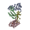


 Mass: 65.409 Da / Num. of mol.: 2 / Source method: obtained synthetically / Formula: Zn
 Mass: 24.305 Da / Num. of mol.: 4 / Source method: obtained synthetically / Formula: Mg
 Mass: 484.141 Da / Num. of mol.: 2 / Source method: obtained synthetically / Formula: C9H15N2O15P3 / Comment: UTP*YM
 Sample preparation
 / Beamline: I02 / Wavelength: 0.9795
  Processing