 ムービー
ムービー コントローラー


+ データを開く

- 基本情報
基本情報
| 登録情報 | データベース: PDB / ID: 2h92 | ||||||
|---|---|---|---|---|---|---|---|
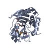
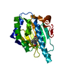
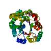
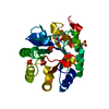
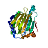
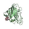
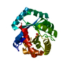
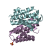
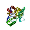
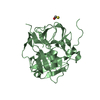
| タイトル | Crystal Structure of Staphylococcus aureus Cytidine Monophosphate Kinase in complex with cytidine-5'-monophosphate | ||||||
 要素 要素 | Cytidylate kinase | ||||||
 キーワード キーワード | TRANSFERASE / Rossmann fold | ||||||
| 機能・相同性 |  機能・相同性情報 機能・相同性情報(d)CMP kinase / CMP kinase activity / dCMP kinase activity / pyrimidine nucleotide metabolic process / nucleobase-containing small molecule interconversion / ATP binding / cytosol 類似検索 - 分子機能 | ||||||
| 生物種 |   Staphylococcus aureus (黄色ブドウ球菌) Staphylococcus aureus (黄色ブドウ球菌) | ||||||
| 手法 |  X線回折 / シンクロトロン / 分子置換 / 解像度: 2.3 Å X線回折 / シンクロトロン / 分子置換 / 解像度: 2.3 Å | ||||||
 データ登録者 データ登録者 | Dhaliwal, B. | ||||||
 引用 引用 | ジャーナル: Acta Crystallogr.,Sect.F / 年: 2006 タイトル: Structure of Staphylococcus aureus cytidine monophosphate kinase in complex with cytidine 5'-monophosphate. 著者: Dhaliwal, B. / Ren, J. / Lockyer, M. / Charles, I. / Hawkins, A.R. / Stammers, D.K. | ||||||
| 履歴 |
|
- 構造の表示
構造の表示
| 構造ビューア | 分子: MolmilJmol/JSmol |
|---|
- ダウンロードとリンク
-ダウンロード
| PDBx/mmCIF形式 | 2h92.cif.gz | 148.1 KB | 表示 | PDBx/mmCIF形式 |
|---|---|---|---|---|
| PDB形式 | pdb2h92.ent.gz | 116.7 KB | 表示 | PDB形式 |
| PDBx/mmJSON形式 | 2h92.json.gz | ツリー表示 | PDBx/mmJSON形式 | |
| その他 |  その他のダウンロード その他のダウンロード |
-検証レポート
| アーカイブディレクトリ | https://data.pdbj.org/pub/pdb/validation_reports/h9/2h92ftp://data.pdbj.org/pub/pdb/validation_reports/h9/2h92 | HTTPS FTP |
|---|
-関連構造データ
| 関連構造データ | 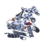 1kdoS S: 精密化の開始モデル |
|---|---|
| 類似構造データ |
-リンク
 PDBj
PDBj- 集合体
集合体
| 登録構造単位 | 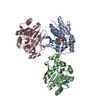
| ||||||||
|---|---|---|---|---|---|---|---|---|---|
| 1 | 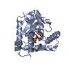
| ||||||||
| 2 | 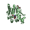
| ||||||||
| 3 | 
| ||||||||
| 単位格子 |
|
-要素
| #1: タンパク質 | 分子量: 24628.996 Da / 分子数: 3 / 由来タイプ: 組換発現 由来: (組換発現) Staphylococcus aureus (黄色ブドウ球菌)遺伝子: cmk / プラスミド: pET15b / 発現宿主: 参照: UniProt: Q1YBX2, UniProt: P63807*PLUS, UMP/CMP kinase #2: 化合物 |   分子量: 96.063 Da / 分子数: 3 / 由来タイプ: 合成 / 式: SO4 分子量: 96.063 Da / 分子数: 3 / 由来タイプ: 合成 / 式: SO4#3: 化合物 |   分子量: 323.197 Da / 分子数: 3 / 由来タイプ: 合成 / 式: C9H14N3O8P 分子量: 323.197 Da / 分子数: 3 / 由来タイプ: 合成 / 式: C9H14N3O8P#4: 化合物 | ChemComp-PG4 / |   分子量: 194.226 Da / 分子数: 1 / 由来タイプ: 合成 / 式: C8H18O5 / コメント: 沈殿剤*YM 分子量: 194.226 Da / 分子数: 1 / 由来タイプ: 合成 / 式: C8H18O5 / コメント: 沈殿剤*YM#5: 水 | ChemComp-HOH / |  分子量: 18.015 Da / 分子数: 334 / 由来タイプ: 天然 / 式: H2O 分子量: 18.015 Da / 分子数: 334 / 由来タイプ: 天然 / 式: H2O |
|---|
-実験情報
-実験
| 実験 | 手法: X線回折 / 使用した結晶の数: 1 |
|---|
- 試料調製
試料調製
| 結晶 | マシュー密度: 3.71 Å3/Da / 溶媒含有率: 66.86 % |
|---|---|
| 結晶化 | 温度: 298 K / 手法: 蒸気拡散法, シッティングドロップ法 / pH: 7.8 詳細: 1.6M ammonium sulfate, 0.1M Hepes, 2% PEG 200, pH 7.8, VAPOR DIFFUSION, SITTING DROP, temperature 298K |
-データ収集
| 回折 | 平均測定温度: 100 K |
|---|---|
| 放射光源 | 由来: シンクロトロン / サイト: ESRF  / ビームライン: ID14-1 / 波長: 0.934 Å / ビームライン: ID14-1 / 波長: 0.934 Å |
| 検出器 | タイプ: ADSC QUANTUM 210 / 検出器: CCD / 日付: 2004年8月5日 |
| 放射 | プロトコル: SINGLE WAVELENGTH / 単色(M)・ラウエ(L): M / 散乱光タイプ: x-ray |
| 放射波長 | 波長: 0.934 Å / 相対比: 1 |
| 反射 | 解像度: 2.3→30 Å / Num. obs: 48287 / % possible obs: 100 % / Observed criterion σ(F): 0 / Observed criterion σ(I): 0 / 冗長度: 13.8 % |
| 反射 シェル | 解像度: 2.3→2.38 Å / 冗長度: 6.9 % / % possible all: 100 |
- 解析
解析
| ソフトウェア |
| ||||||||||||||||||||
|---|---|---|---|---|---|---|---|---|---|---|---|---|---|---|---|---|---|---|---|---|---|
| 精密化 | 構造決定の手法: 分子置換 開始モデル: PDB ENTRY 1KDO 解像度: 2.3→30 Å / 交差検証法: THROUGHOUT / σ(F): 0 / 立体化学のターゲット値: Engh & Huber
| ||||||||||||||||||||
| 精密化ステップ | サイクル: LAST / 解像度: 2.3→30 Å
| ||||||||||||||||||||
| 拘束条件 |
| ||||||||||||||||||||
| Xplor file |
|
