 Movie
Movie Controller


+ Open data

- Basic information
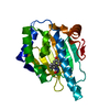
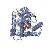
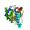
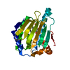
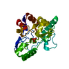
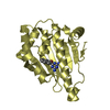
| Entry | Database: PDB / ID: 5v44 | ||||||
|---|---|---|---|---|---|---|---|
| Title | Crystal structure of the SR1 domain of human sacsin | ||||||
 Components Components | Sacsin | ||||||
 Keywords Keywords | CHAPERONE / alpha-beta sandwich / Bergerat fold | ||||||
| Function / homology |  Function and homology information Function and homology informationnegative regulation of inclusion body assembly / cell body fiber / low-density lipoprotein particle receptor binding / proteasome binding / Hsp70 protein binding / protein-folding chaperone binding / protein folding / axon / dendrite / mitochondrion ...negative regulation of inclusion body assembly / cell body fiber / low-density lipoprotein particle receptor binding / proteasome binding / Hsp70 protein binding / protein-folding chaperone binding / protein folding / axon / dendrite / mitochondrion / identical protein binding / nucleus / cytoplasm Similarity search - Function | ||||||
| Biological species |  Homo sapiens (human) Homo sapiens (human) | ||||||
| Method |  X-RAY DIFFRACTION / SYNCHROTRON / MOLECULAR REPLACEMENT / Resolution: 1.56 Å X-RAY DIFFRACTION / SYNCHROTRON / MOLECULAR REPLACEMENT / Resolution: 1.56 Å | ||||||
 Authors Authors | Menade, M. / Kozlov, G. / Gehring, K. | ||||||
| Funding support |  Canada, 1items Canada, 1items
| ||||||
 Citation Citation | Journal: J. Biol. Chem. / Year: 2018 Title: Structures of ubiquitin-like (Ubl) and Hsp90-like domains of sacsin provide insight into pathological mutations. Authors: Menade, M. / Kozlov, G. / Trempe, J.F. / Pande, H. / Shenker, S. / Wickremasinghe, S. / Li, X. / Hojjat, H. / Dicaire, M.J. / Brais, B. / McPherson, P.S. / Wong, M.J.H. / Young, J.C. / Gehring, K. | ||||||
| History |
|
- Structure visualization
| Structure viewer | Molecule: MolmilJmol/JSmol |
|---|
- Downloads & links
-Download
| PDBx/mmCIF format | 5v44.cif.gz | 295.8 KB | Display | PDBx/mmCIF format |
|---|---|---|---|---|
| PDB format | pdb5v44.ent.gz | 240.9 KB | Display | PDB format |
| PDBx/mmJSON format | 5v44.json.gz | Tree view | PDBx/mmJSON format | |
| Others |  Other downloads Other downloads |
-Validation report
| Arichive directory | https://data.pdbj.org/pub/pdb/validation_reports/v4/5v44ftp://data.pdbj.org/pub/pdb/validation_reports/v4/5v44 | HTTPS FTP |
|---|
-Related structure data
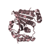
| Related structure data | 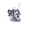 5v45SC 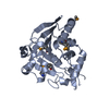 5v46C 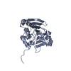 5v47C  5vsxC  5vszC S: Starting model for refinement C: citing same article ( |
|---|---|
| Similar structure data |

-Links
 PDBj
PDBj


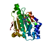
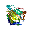
- Assembly
| Deposited unit | 
| ||||||||
|---|---|---|---|---|---|---|---|---|---|
| 1 | 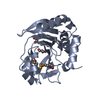
| ||||||||
| 2 | 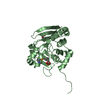
| ||||||||
| 3 | 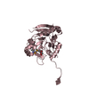
| ||||||||
| Unit cell |
|
-Components
| #1: Protein | Mass: 28636.795 Da / Num. of mol.: 3 Source method: isolated from a genetically manipulated source Source: (gene. exp.) Homo sapiens (human) / Gene: SACS, KIAA0730 / Plasmid: pGEX-6P-1 / Production host:  #2: Chemical | ChemComp-GOL /   Mass: 92.094 Da / Num. of mol.: 6 / Source method: obtained synthetically / Formula: C3H8O3 Mass: 92.094 Da / Num. of mol.: 6 / Source method: obtained synthetically / Formula: C3H8O3#3: Water | ChemComp-HOH / |  Mass: 18.015 Da / Num. of mol.: 389 / Source method: isolated from a natural source / Formula: H2O Mass: 18.015 Da / Num. of mol.: 389 / Source method: isolated from a natural source / Formula: H2OHas protein modification | Y | |
|---|
-Experimental details
-Experiment
| Experiment | Method: X-RAY DIFFRACTION / Number of used crystals: 1 |
|---|
- Sample preparation
| Crystal | Density Matthews: 1.98 Å3/Da / Density % sol: 37.97 % |
|---|---|
| Crystal grow | Temperature: 293 K / Method: vapor diffusion, hanging drop / pH: 6.5 Details: 28% PEG 3350, 0.2 M ammonium acetate, 0.1 M MES pH 6.5 |
-Data collection
| Diffraction | Mean temperature: 100 K |
|---|---|
| Diffraction source | Source: SYNCHROTRON / Site: CHESS  / Beamline: A1 / Wavelength: 0.977 Å / Beamline: A1 / Wavelength: 0.977 Å |
| Detector | Type: ADSC QUANTUM 210 / Detector: CCD / Date: Mar 12, 2014 |
| Radiation | Protocol: SINGLE WAVELENGTH / Monochromatic (M) / Laue (L): M / Scattering type: x-ray |
| Radiation wavelength | Wavelength: 0.977 Å / Relative weight: 1 |
| Reflection | Resolution: 1.55→50 Å / Num. obs: 86247 / % possible obs: 96.25 % / Redundancy: 4.3 % / Rsym value: 0.049 / Net I/σ(I): 34.6 |
| Reflection shell | Resolution: 1.55→1.59 Å / Redundancy: 4.2 % / Mean I/σ(I) obs: 2.9 / Num. unique obs: 5846 / Rsym value: 0.449 / % possible all: 87.96 |
- Processing
| Software |
| ||||||||||||||||||||||||||||||||||||||||||||||||||||||||||||||||||||||||||||||||||||||||||||||||||||||||||||||||||||||||||||||||||||||||||||||||||||||||||||||||||||||||||||||||||||||
|---|---|---|---|---|---|---|---|---|---|---|---|---|---|---|---|---|---|---|---|---|---|---|---|---|---|---|---|---|---|---|---|---|---|---|---|---|---|---|---|---|---|---|---|---|---|---|---|---|---|---|---|---|---|---|---|---|---|---|---|---|---|---|---|---|---|---|---|---|---|---|---|---|---|---|---|---|---|---|---|---|---|---|---|---|---|---|---|---|---|---|---|---|---|---|---|---|---|---|---|---|---|---|---|---|---|---|---|---|---|---|---|---|---|---|---|---|---|---|---|---|---|---|---|---|---|---|---|---|---|---|---|---|---|---|---|---|---|---|---|---|---|---|---|---|---|---|---|---|---|---|---|---|---|---|---|---|---|---|---|---|---|---|---|---|---|---|---|---|---|---|---|---|---|---|---|---|---|---|---|---|---|---|---|
| Refinement | Method to determine structure: MOLECULAR REPLACEMENT Starting model: 5V45 Resolution: 1.56→50 Å / Cor.coef. Fo:Fc: 0.963 / Cor.coef. Fo:Fc free: 0.945 / SU B: 3.229 / SU ML: 0.058 / Cross valid method: THROUGHOUT / ESU R: 0.091 / ESU R Free: 0.091
| ||||||||||||||||||||||||||||||||||||||||||||||||||||||||||||||||||||||||||||||||||||||||||||||||||||||||||||||||||||||||||||||||||||||||||||||||||||||||||||||||||||||||||||||||||||||
| Solvent computation | Ion probe radii: 0.8 Å / Shrinkage radii: 0.8 Å / VDW probe radii: 1.2 Å | ||||||||||||||||||||||||||||||||||||||||||||||||||||||||||||||||||||||||||||||||||||||||||||||||||||||||||||||||||||||||||||||||||||||||||||||||||||||||||||||||||||||||||||||||||||||
| Displacement parameters | Biso mean: 31.172 Å2
| ||||||||||||||||||||||||||||||||||||||||||||||||||||||||||||||||||||||||||||||||||||||||||||||||||||||||||||||||||||||||||||||||||||||||||||||||||||||||||||||||||||||||||||||||||||||
| Refinement step | Cycle: 1 / Resolution: 1.56→50 Å
| ||||||||||||||||||||||||||||||||||||||||||||||||||||||||||||||||||||||||||||||||||||||||||||||||||||||||||||||||||||||||||||||||||||||||||||||||||||||||||||||||||||||||||||||||||||||
| Refine LS restraints |
|