| Entry | Database: PDB / ID: 5v47
|
|---|
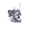
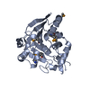
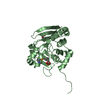
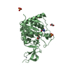
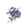
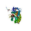
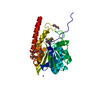
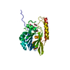
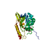
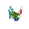
| Title | Crystal structure of the SR1 domain of lizard sacsin |
|---|
 Components Components | Lizard sacsin |
|---|
 Keywords Keywords | CHAPERONE / alpha-beta sandwich / Bergerat fold |
|---|
| Function / homology |  Function and homology information Function and homology information
: / HEPN domain profile. / Higher Eukarytoes and Prokaryotes Nucleotide-binding domain / HEPN domain / HEPN domain / dnaJ domain profile. / Chaperone J-domain superfamily / DnaJ domain / Histidine kinase/HSP90-like ATPase superfamily / Ubiquitin family ...: / HEPN domain profile. / Higher Eukarytoes and Prokaryotes Nucleotide-binding domain / HEPN domain / HEPN domain / dnaJ domain profile. / Chaperone J-domain superfamily / DnaJ domain / Histidine kinase/HSP90-like ATPase superfamily / Ubiquitin family / Ubiquitin homologues / Ubiquitin domain profile. / Ubiquitin-like domain / Ubiquitin-like domain superfamilySimilarity search - Domain/homology |
|---|
| Biological species |  Anolis carolinensis (green anole) Anolis carolinensis (green anole) |
|---|
| Method |  X-RAY DIFFRACTION / SYNCHROTRON / MOLECULAR REPLACEMENT / Resolution: 1.84 Å X-RAY DIFFRACTION / SYNCHROTRON / MOLECULAR REPLACEMENT / Resolution: 1.84 Å |
|---|
 Authors Authors | Pan, T. / Menade, M. / Kozlov, G. / Gehring, K. |
|---|
| Funding support |  Canada, 1items Canada, 1items | Organization | Grant number | Country |
|---|
| ARSACS Foundation | | Canada |
|
|---|
 Citation Citation | Journal: J. Biol. Chem. / Year: 2018
Title: Structures of ubiquitin-like (Ubl) and Hsp90-like domains of sacsin provide insight into pathological mutations.
Authors: Menade, M. / Kozlov, G. / Trempe, J.F. / Pande, H. / Shenker, S. / Wickremasinghe, S. / Li, X. / Hojjat, H. / Dicaire, M.J. / Brais, B. / McPherson, P.S. / Wong, M.J.H. / Young, J.C. / Gehring, K. |
|---|
| History | | Deposition | Mar 8, 2017 | Deposition site: RCSB / Processing site: RCSB |
|---|
| Revision 1.0 | May 24, 2017 | Provider: repository / Type: Initial release |
|---|
| Revision 1.1 | Jul 11, 2018 | Group: Data collection / Database references / Category: citation / citation_author
Item: _citation.country / _citation.journal_abbrev ..._citation.country / _citation.journal_abbrev / _citation.journal_id_ASTM / _citation.journal_id_CSD / _citation.journal_id_ISSN / _citation.pdbx_database_id_DOI / _citation.pdbx_database_id_PubMed / _citation.title / _citation.year |
|---|
| Revision 1.2 | Aug 29, 2018 | Group: Data collection / Database references / Category: citation / citation_author
Item: _citation.journal_volume / _citation.page_first ..._citation.journal_volume / _citation.page_first / _citation.page_last / _citation.title / _citation_author.identifier_ORCID |
|---|
| Revision 1.3 | Oct 4, 2023 | Group: Data collection / Database references / Refinement description
Category: chem_comp_atom / chem_comp_bond ...chem_comp_atom / chem_comp_bond / database_2 / pdbx_initial_refinement_model
Item: _database_2.pdbx_DOI / _database_2.pdbx_database_accession |
|---|
|
|---|
 Movie
Movie Controller


 Open data
 Basic information
 Structure visualization
 Downloads & links
 Other downloads
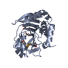


 PDBj
PDBj


 Assembly


 Mass: 96.063 Da / Num. of mol.: 1 / Source method: obtained synthetically / Formula: SO4
 Mass: 18.015 Da / Num. of mol.: 252 / Source method: isolated from a natural source / Formula: H2O
 Sample preparation
 / Beamline: F1 / Wavelength: 0.977 Å
 Processing