Mass: 18.015 Da / Num. of mol.: 295 / Source method: isolated from a natural source / Formula: H2O
-
Experimental details
-
Experiment
Experiment
Method: X-RAY DIFFRACTION
-
Sample preparation
Crystal
Density Matthews: 2.41 Å3/Da / Density % sol: 48.92 %
Crystal grow
Temperature: 289 K / Method: vapor diffusion, sitting drop Details: 0.2 M Li2SO4, 0.1 M CAPS:NaOH pH 10.5, 1.2 M/NaH2PO4, 0.8 M K2HPO4, cryo 2.4 M K2HPO4
Resolution: 1.05→28.531 Å / SU ML: 0.08 / Cross valid method: THROUGHOUT / σ(F): 1.36 / Phase error: 12.49 / Stereochemistry target values: ML / Details: Hydrogen atoms have been added in riding positions
Rfactor
Num. reflection
% reflection
Selection details
Rfree
0.141
1145
1 %
random
Rwork
0.1226
-
-
-
obs
0.1228
114252
99.43 %
-
Solvent computation
Shrinkage radii: 0.9 Å / VDW probe radii: 1.11 Å / Solvent model: FLAT BULK SOLVENT MODEL
Refinement step
Cycle: LAST / Resolution: 1.05→28.531 Å
Protein
Nucleic acid
Ligand
Solvent
Total
Num. atoms
1766
0
34
295
2095
Refine LS restraints
Refine-ID
Type
Dev ideal
Number
X-RAY DIFFRACTION
f_bond_d
0.014
1978
X-RAY DIFFRACTION
f_angle_d
1.603
2718
X-RAY DIFFRACTION
f_dihedral_angle_d
13.315
720
X-RAY DIFFRACTION
f_chiral_restr
0.101
318
X-RAY DIFFRACTION
f_plane_restr
0.009
364
LS refinement shell
Resolution (Å)
Rfactor Rfree
Num. reflection Rfree
Rfactor Rwork
Num. reflection Rwork
Refine-ID
% reflection obs (%)
1.0501-1.0979
0.2212
151
0.1909
13578
X-RAY DIFFRACTION
96
1.0979-1.1558
0.1446
132
0.1312
14172
X-RAY DIFFRACTION
100
1.1558-1.2282
0.1078
162
0.1007
14143
X-RAY DIFFRACTION
100
1.2282-1.323
0.1288
154
0.0904
14128
X-RAY DIFFRACTION
100
1.323-1.4561
0.1062
128
0.0852
14204
X-RAY DIFFRACTION
100
1.4561-1.6668
0.109
146
0.0876
14231
X-RAY DIFFRACTION
100
1.6668-2.0999
0.1126
138
0.1192
14259
X-RAY DIFFRACTION
100
2.0999-28.5411
0.1726
134
0.1399
14392
X-RAY DIFFRACTION
99
+
About Yorodumi
-
News
-
Feb 9, 2022. New format data for meta-information of EMDB entries
New format data for meta-information of EMDB entries
Version 3 of the EMDB header file is now the official format.
The previous official version 1.9 will be removed from the archive.
In the structure databanks used in Yorodumi, some data are registered as the other names, "COVID-19 virus" and "2019-nCoV". Here are the details of the virus and the list of structure data.
Jan 31, 2019. EMDB accession codes are about to change! (news from PDBe EMDB page)
EMDB accession codes are about to change! (news from PDBe EMDB page)
The allocation of 4 digits for EMDB accession codes will soon come to an end. Whilst these codes will remain in use, new EMDB accession codes will include an additional digit and will expand incrementally as the available range of codes is exhausted. The current 4-digit format prefixed with “EMD-” (i.e. EMD-XXXX) will advance to a 5-digit format (i.e. EMD-XXXXX), and so on. It is currently estimated that the 4-digit codes will be depleted around Spring 2019, at which point the 5-digit format will come into force.
The EM Navigator/Yorodumi systems omit the EMD- prefix.
Related info.:Q: What is EMD? / ID/Accession-code notation in Yorodumi/EM Navigator
Yorodumi is a browser for structure data from EMDB, PDB, SASBDB, etc.
This page is also the successor to EM Navigator detail page, and also detail information page/front-end page for Omokage search.
The word "yorodu" (or yorozu) is an old Japanese word meaning "ten thousand". "mi" (miru) is to see.
Related info.:EMDB / PDB / SASBDB / Comparison of 3 databanks / Yorodumi Search / Aug 31, 2016. New EM Navigator & Yorodumi / Yorodumi Papers / Jmol/JSmol / Function and homology information / Changes in new EM Navigator and Yorodumi
 Movie
Movie Controller


 Open data
 Basic information
 Components
 Keywords
 Function and homology information
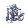
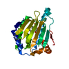
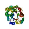
 Actinomyces urogenitalis DSM 15434 (bacteria)
 X-RAY DIFFRACTION /
  Authors
 United States, 1items
  Citation
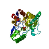
 Structure visualization
 Downloads & links
 Other downloads
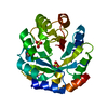


 PDBj
PDBj
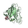
 Assembly
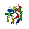

 Mass: 94.971 Da / Num. of mol.: 4 / Source method: obtained synthetically / Formula: PO4
 Mass: 221.317 Da / Num. of mol.: 1 / Source method: obtained synthetically / Formula: C9H19NO3S / Comment: pH buffer*YM
 Mass: 18.015 Da / Num. of mol.: 295 / Source method: isolated from a natural source / Formula: H2O
 Sample preparation
 Processing