 Movie
Movie Controller

[English] 日本語
 Yorodumi
- PDB-3wyz: On archaeal homologs of the human RNase P protein Rpp30 in the hy... -
+ Open data

- Basic information
| Entry | Database: PDB / ID: 3wyz | ||||||
|---|---|---|---|---|---|---|---|
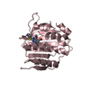
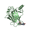
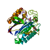
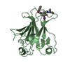
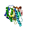
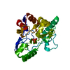
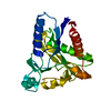
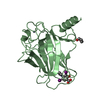
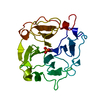
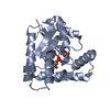
| Title | On archaeal homologs of the human RNase P protein Rpp30 in the hyperthermophilic archaeon Thermococcus kodakarensis | ||||||
 Components Components | Ribonuclease P protein component 3 | ||||||
 Keywords Keywords | HYDROLASE / TIM barrel-like structure / pre-tRNA cleavage / RNA binding | ||||||
| Function / homology |  Function and homology information Function and homology informationribonuclease P complex / ribonuclease P / ribonuclease P activity / tRNA 5'-leader removal / RNA binding / cytoplasm Similarity search - Function | ||||||
| Biological species |   Thermococcus kodakarensis KOD1 (archaea) Thermococcus kodakarensis KOD1 (archaea) | ||||||
| Method |  X-RAY DIFFRACTION / SYNCHROTRON / MOLECULAR REPLACEMENT / Resolution: 2.21 Å X-RAY DIFFRACTION / SYNCHROTRON / MOLECULAR REPLACEMENT / Resolution: 2.21 Å | ||||||
 Authors Authors | Suematsu, K. / Ueda, T. / Nakashima, T. / Kakuta, Y. / Kimura, M. | ||||||
 Citation Citation | Journal: Biosci.Biotechnol.Biochem. / Year: 2015 Title: On archaeal homologs of the human RNase P proteins Pop5 and Rpp30 in the hyperthermophilic archaeon Thermococcus kodakarensis. Authors: Suematsu, K. / Ueda, T. / Nakashima, T. / Kakuta, Y. / Kimura, M. | ||||||
| History |
|
- Structure visualization
| Structure viewer | Molecule: MolmilJmol/JSmol |
|---|
- Downloads & links
-Download
| PDBx/mmCIF format | 3wyz.cif.gz | 55.3 KB | Display | PDBx/mmCIF format |
|---|---|---|---|---|
| PDB format | pdb3wyz.ent.gz | 40.2 KB | Display | PDB format |
| PDBx/mmJSON format | 3wyz.json.gz | Tree view | PDBx/mmJSON format | |
| Others |  Other downloads Other downloads |
-Validation report
| Arichive directory | https://data.pdbj.org/pub/pdb/validation_reports/wy/3wyzftp://data.pdbj.org/pub/pdb/validation_reports/wy/3wyz | HTTPS FTP |
|---|
-Related structure data

-Links
 PDBj
PDBj
- Assembly
| Deposited unit | 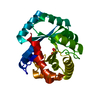
| ||||||||
|---|---|---|---|---|---|---|---|---|---|
| 1 |
| ||||||||
| Unit cell |
|
-Components
| #1: Protein | Mass: 25306.330 Da / Num. of mol.: 1 Source method: isolated from a genetically manipulated source Source: (gene. exp.) Thermococcus kodakarensis KOD1 (archaea)Gene: rnp3, TK1450 / Production host:  |
|---|---|
| #2: Chemical | ChemComp-GOL /   Mass: 92.094 Da / Num. of mol.: 1 / Source method: obtained synthetically / Formula: C3H8O3 Mass: 92.094 Da / Num. of mol.: 1 / Source method: obtained synthetically / Formula: C3H8O3 |
| #3: Water | ChemComp-HOH /  Mass: 18.015 Da / Num. of mol.: 39 / Source method: isolated from a natural source / Formula: H2O Mass: 18.015 Da / Num. of mol.: 39 / Source method: isolated from a natural source / Formula: H2O |
-Experimental details
-Experiment
| Experiment | Method: X-RAY DIFFRACTION / Number of used crystals: 1 |
|---|
- Sample preparation
| Crystal | Density Matthews: 1.92 Å3/Da / Density % sol: 35.95 % |
|---|---|
| Crystal grow | Temperature: 283 K / Method: purification / pH: 7.5 Details: 50mM Tris-HCl (pH 7.5), 200mM NaCl, 10mM MgCl2, Purification, temperature 283K |
-Data collection
| Diffraction | Mean temperature: 100 K |
|---|---|
| Diffraction source | Source: SYNCHROTRON / Site: SPring-8  / Beamline: BL44XU / Wavelength: 0.9 Å / Beamline: BL44XU / Wavelength: 0.9 Å |
| Detector | Type: RAYONIX MX300HE / Detector: CCD |
| Radiation | Protocol: SINGLE WAVELENGTH / Monochromatic (M) / Laue (L): M / Scattering type: x-ray |
| Radiation wavelength | Wavelength: 0.9 Å / Relative weight: 1 |
| Reflection | Resolution: 2.2→50 Å / Num. all: 9422 / Num. obs: 9422 / % possible obs: 96.8 % / Observed criterion σ(F): 0 / Observed criterion σ(I): 0 / Redundancy: 3.2 % / Rmerge(I) obs: 0.141 / Net I/σ(I): 18.4 |
| Reflection shell | Resolution: 2.2→2.3 Å / Redundancy: 3 % / Rmerge(I) obs: 0.917 / Mean I/σ(I) obs: 2.7 / % possible all: 93.5 |
- Processing
| Software |
| |||||||||||||||||||||||||||||||||||||||||||||||||||||||||||||||||||||||||||||||||||||||||||||||||||||||||
|---|---|---|---|---|---|---|---|---|---|---|---|---|---|---|---|---|---|---|---|---|---|---|---|---|---|---|---|---|---|---|---|---|---|---|---|---|---|---|---|---|---|---|---|---|---|---|---|---|---|---|---|---|---|---|---|---|---|---|---|---|---|---|---|---|---|---|---|---|---|---|---|---|---|---|---|---|---|---|---|---|---|---|---|---|---|---|---|---|---|---|---|---|---|---|---|---|---|---|---|---|---|---|---|---|---|---|
| Refinement | Method to determine structure: MOLECULAR REPLACEMENT / Resolution: 2.21→33.48 Å / Cor.coef. Fo:Fc: 0.953 / Cor.coef. Fo:Fc free: 0.925 / SU B: 10.894 / SU ML: 0.262 / Cross valid method: THROUGHOUT / σ(F): 0 / σ(I): 0 / ESU R: 0.51 / ESU R Free: 0.288 / Stereochemistry target values: MAXIMUM LIKELIHOOD / Details: HYDROGENS HAVE BEEN ADDED IN THE RIDING POSITIONS
| |||||||||||||||||||||||||||||||||||||||||||||||||||||||||||||||||||||||||||||||||||||||||||||||||||||||||
| Solvent computation | Ion probe radii: 0.8 Å / Shrinkage radii: 0.8 Å / VDW probe radii: 1.2 Å / Solvent model: MASK | |||||||||||||||||||||||||||||||||||||||||||||||||||||||||||||||||||||||||||||||||||||||||||||||||||||||||
| Displacement parameters | Biso mean: 56.06 Å2
| |||||||||||||||||||||||||||||||||||||||||||||||||||||||||||||||||||||||||||||||||||||||||||||||||||||||||
| Refinement step | Cycle: LAST / Resolution: 2.21→33.48 Å
| |||||||||||||||||||||||||||||||||||||||||||||||||||||||||||||||||||||||||||||||||||||||||||||||||||||||||
| Refine LS restraints |
| |||||||||||||||||||||||||||||||||||||||||||||||||||||||||||||||||||||||||||||||||||||||||||||||||||||||||
| LS refinement shell | Resolution: 2.206→2.263 Å / Total num. of bins used: 20
|
