 Movie
Movie Controller

[English] 日本語
 Yorodumi
- PDB-1sfr: Crystal Structure of the Mycobacterium tuberculosis Antigen 85A P... -
+ Open data

- Basic information
| Entry | Database: PDB / ID: 1sfr | ||||||
|---|---|---|---|---|---|---|---|
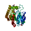
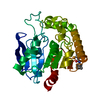
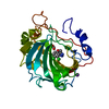
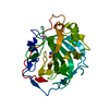
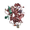
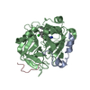
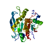
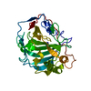
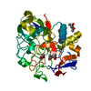
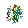
| Title | Crystal Structure of the Mycobacterium tuberculosis Antigen 85A Protein | ||||||
 Components Components | Antigen 85-A | ||||||
 Keywords Keywords | TRANSFERASE / alpha/beta hydrolase / Structural Genomics / PSI / Protein Structure Initiative / TB Structural Genomics Consortium / TBSGC | ||||||
| Function / homology |  Function and homology information Function and homology informationtrehalose O-mycolyltransferase activity / trehalose O-mycolyltransferase / diacylglycerol O-acyltransferase / diacylglycerol O-acyltransferase activity / cell wall / zymogen binding / acyltransferase activity, transferring groups other than amino-acyl groups / peptidoglycan-based cell wall / extracellular region / plasma membrane / cytoplasm Similarity search - Function | ||||||
| Biological species |   Mycobacterium tuberculosis (bacteria) Mycobacterium tuberculosis (bacteria) | ||||||
| Method |  X-RAY DIFFRACTION / SYNCHROTRON / MOLECULAR REPLACEMENT / Resolution: 2.7 Å X-RAY DIFFRACTION / SYNCHROTRON / MOLECULAR REPLACEMENT / Resolution: 2.7 Å | ||||||
 Authors Authors | Ronning, D.R. / Vissa, V. / Besra, G.S. / Belisle, J.T. / Sacchettini, J.C. / TB Structural Genomics Consortium (TBSGC) | ||||||
 Citation Citation | Journal: J.Biol.Chem. / Year: 2004 Title: Mycobacterium tuberculosis Antigen 85A and 85C Structures Confirm Binding Orientation and Conserved Substrate Specificity Authors: Ronning, D.R. / Vissa, V. / Besra, G.S. / Belisle, J.T. / Sacchettini, J.C. | ||||||
| History |
|
- Structure visualization
| Structure viewer | Molecule: MolmilJmol/JSmol |
|---|
- Downloads & links
-Download
| PDBx/mmCIF format | 1sfr.cif.gz | 167.5 KB | Display | PDBx/mmCIF format |
|---|---|---|---|---|
| PDB format | pdb1sfr.ent.gz | 134.8 KB | Display | PDB format |
| PDBx/mmJSON format | 1sfr.json.gz | Tree view | PDBx/mmJSON format | |
| Others |  Other downloads Other downloads |
-Validation report
| Arichive directory | https://data.pdbj.org/pub/pdb/validation_reports/sf/1sfrftp://data.pdbj.org/pub/pdb/validation_reports/sf/1sfr | HTTPS FTP |
|---|
-Related structure data
| Related structure data |  1va5C  1dqzS S: Starting model for refinement C: citing same article ( |
|---|---|
| Similar structure data | |
| Other databases |


-Links
 PDBj
PDBj

- Assembly
| Deposited unit | 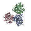
| ||||||||
|---|---|---|---|---|---|---|---|---|---|
| 1 | 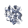
| ||||||||
| 2 | 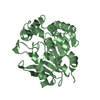
| ||||||||
| 3 | 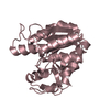
| ||||||||
| 4 | 
| ||||||||
| 5 | 
| ||||||||
| Unit cell |
| ||||||||
| Details | The biological assembly is a monomer |
-Components
| #1: Protein | Mass: 32744.457 Da / Num. of mol.: 3 Source method: isolated from a genetically manipulated source Source: (gene. exp.) Mycobacterium tuberculosis (bacteria) / Gene: fbpA / Plasmid: pET30b / Species (production host): Escherichia coli / Production host: References: UniProt: P0A4V2, UniProt: P9WQP3*PLUS, Transferases; Acyltransferases; Transferring groups other than aminoacyl groups #2: Water | ChemComp-HOH / |  Mass: 18.015 Da / Num. of mol.: 58 / Source method: isolated from a natural source / Formula: H2O Mass: 18.015 Da / Num. of mol.: 58 / Source method: isolated from a natural source / Formula: H2OHas protein modification | Y | |
|---|
-Experimental details
-Experiment
| Experiment | Method: X-RAY DIFFRACTION / Number of used crystals: 1 |
|---|
- Sample preparation
| Crystal | Density Matthews: 5.23 Å3/Da / Density % sol: 76.31 % |
|---|---|
| Crystal grow | Temperature: 293 K / Method: vapor diffusion, hanging drop / pH: 6.5 Details: Sodium Cacodylate, Sodium Acetate, pH 6.5, VAPOR DIFFUSION, HANGING DROP, temperature 293K |
-Data collection
| Diffraction | Mean temperature: 100 K |
|---|---|
| Diffraction source | Source: SYNCHROTRON / Site: APS  / Beamline: 14-BM-C / Wavelength: 1 Å / Beamline: 14-BM-C / Wavelength: 1 Å |
| Detector | Type: ADSC QUANTUM 4 / Detector: CCD / Date: Oct 14, 2000 |
| Radiation | Monochromator: Ge 111 / Protocol: SINGLE WAVELENGTH / Monochromatic (M) / Laue (L): M / Scattering type: x-ray |
| Radiation wavelength | Wavelength: 1 Å / Relative weight: 1 |
| Reflection | Resolution: 2.7→30 Å / Num. all: 52327 / Num. obs: 52327 / % possible obs: 98.5 % / Observed criterion σ(F): 0 / Observed criterion σ(I): 0 / Redundancy: 4 % / Biso Wilson estimate: 59.1 Å2 / Rsym value: 0.069 / Net I/σ(I): 15 |
| Reflection shell | Resolution: 2.7→2.8 Å / % possible all: 98 |
- Processing
| Software |
| ||||||||||||||||||||||||||||||||||||
|---|---|---|---|---|---|---|---|---|---|---|---|---|---|---|---|---|---|---|---|---|---|---|---|---|---|---|---|---|---|---|---|---|---|---|---|---|---|
| Refinement | Method to determine structure: MOLECULAR REPLACEMENT Starting model: PDB ENTRY 1DQZ Resolution: 2.7→29.9 Å / Rfactor Rfree error: 0.005 / Data cutoff high absF: 481759.2 / Data cutoff low absF: 0 / Isotropic thermal model: RESTRAINED / Cross valid method: THROUGHOUT / σ(F): 0 / Stereochemistry target values: Engh & Huber
| ||||||||||||||||||||||||||||||||||||
| Solvent computation | Solvent model: FLAT MODEL / Bsol: 35.2765 Å2 / ksol: 0.354084 e/Å3 | ||||||||||||||||||||||||||||||||||||
| Displacement parameters | Biso mean: 54.2 Å2
| ||||||||||||||||||||||||||||||||||||
| Refine analyze |
| ||||||||||||||||||||||||||||||||||||
| Refinement step | Cycle: LAST / Resolution: 2.7→29.9 Å
| ||||||||||||||||||||||||||||||||||||
| Refine LS restraints |
| ||||||||||||||||||||||||||||||||||||
| LS refinement shell | Resolution: 2.7→2.87 Å / Rfactor Rfree error: 0.017 / Total num. of bins used: 6
| ||||||||||||||||||||||||||||||||||||
| Xplor file |
|
