 Movie
Movie Controller


+ Open data

- Basic information
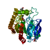
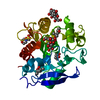
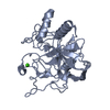
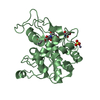
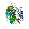
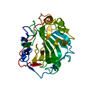
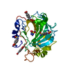
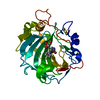
| Entry | Database: PDB / ID: 1f0n | ||||||
|---|---|---|---|---|---|---|---|
| Title | MYCOBACTERIUM TUBERCULOSIS ANTIGEN 85B | ||||||
 Components Components | ANTIGEN 85B | ||||||
 Keywords Keywords | TRANSFERASE / MYCOLYL TRANSFERASE / 30KDA SECRETORY PROTEIN / ANTIGEN 85B / Structural Genomics / PSI / Protein Structure Initiative / TB Structural Genomics Consortium / TBSGC | ||||||
| Function / homology |  Function and homology information Function and homology informationtrehalose O-mycolyltransferase activity / trehalose O-mycolyltransferase / diacylglycerol O-acyltransferase / diacylglycerol O-acyltransferase activity / regulation of growth rate / mycolate cell wall layer assembly / positive regulation of plasminogen activation / cell wall / response to host immune response / zymogen binding ...trehalose O-mycolyltransferase activity / trehalose O-mycolyltransferase / diacylglycerol O-acyltransferase / diacylglycerol O-acyltransferase activity / regulation of growth rate / mycolate cell wall layer assembly / positive regulation of plasminogen activation / cell wall / response to host immune response / zymogen binding / acyltransferase activity, transferring groups other than amino-acyl groups / fibronectin binding / peptidoglycan-based cell wall / response to antibiotic / extracellular region / plasma membrane Similarity search - Function | ||||||
| Biological species |   Mycobacterium tuberculosis (bacteria) Mycobacterium tuberculosis (bacteria) | ||||||
| Method |  X-RAY DIFFRACTION / SYNCHROTRON / MOLECULAR REPLACEMENT / Resolution: 1.8 Å X-RAY DIFFRACTION / SYNCHROTRON / MOLECULAR REPLACEMENT / Resolution: 1.8 Å | ||||||
 Authors Authors | Anderson, D.H. / Harth, G. / Horwitz, M.A. / Eisenberg, D. / TB Structural Genomics Consortium (TBSGC) | ||||||
 Citation Citation | Journal: J.Mol.Biol. / Year: 2001 Title: An interfacial mechanism and a class of inhibitors inferred from two crystal structures of the Mycobacterium tuberculosis 30 kDa major secretory protein (Antigen 85B), a mycolyl transferase. Authors: Anderson, D.H. / Harth, G. / Horwitz, M.A. / Eisenberg, D. #1: Journal: Nat.Struct.Biol. / Year: 2000Title: Crystal Structure of the Secreted Form of Antigen 85C Reveals Potential Targets for Mycobacterial Drugs and Vaccines Authors: Ronning, D.R. / Klabunde, T. / Besra, G.S. / Vissa, V.D. / Belisle, J.T. / Sacchettini, J.C. | ||||||
| History |
|
- Structure visualization
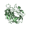
| Structure viewer | Molecule: MolmilJmol/JSmol |
|---|
- Downloads & links
-Download
| PDBx/mmCIF format | 1f0n.cif.gz | 117.3 KB | Display | PDBx/mmCIF format |
|---|---|---|---|---|
| PDB format | pdb1f0n.ent.gz | 90.9 KB | Display | PDB format |
| PDBx/mmJSON format | 1f0n.json.gz | Tree view | PDBx/mmJSON format | |
| Others |  Other downloads Other downloads |
-Validation report
| Arichive directory | https://data.pdbj.org/pub/pdb/validation_reports/f0/1f0nftp://data.pdbj.org/pub/pdb/validation_reports/f0/1f0n | HTTPS FTP |
|---|
-Related structure data


-Links
 PDBj
PDBj- Assembly
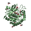
| Deposited unit | 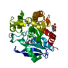
| ||||||||
|---|---|---|---|---|---|---|---|---|---|
| 1 |
| ||||||||
| Unit cell |
| ||||||||
| Components on special symmetry positions |
|
-Components
| #1: Protein | Mass: 30680.012 Da / Num. of mol.: 1 Source method: isolated from a genetically manipulated source Source: (gene. exp.) Mycobacterium tuberculosis (bacteria) / Plasmid: PSMT3 / Production host: Mycobacterium smegmatis (bacteria) / References: UniProt: P31952, UniProt: P9WQP1*PLUS | ||||
|---|---|---|---|---|---|
| #2: Chemical | ChemComp-MES /   Mass: 195.237 Da / Num. of mol.: 1 / Source method: obtained synthetically / Formula: C6H13NO4S / Comment: pH buffer*YM Mass: 195.237 Da / Num. of mol.: 1 / Source method: obtained synthetically / Formula: C6H13NO4S / Comment: pH buffer*YM | ||||
| #3: Chemical |   Mass: 118.174 Da / Num. of mol.: 3 / Source method: obtained synthetically / Formula: C6H14O2 / Comment: precipitant*YM Mass: 118.174 Da / Num. of mol.: 3 / Source method: obtained synthetically / Formula: C6H14O2 / Comment: precipitant*YM#4: Water | ChemComp-HOH / |  Mass: 18.015 Da / Num. of mol.: 205 / Source method: isolated from a natural source / Formula: H2O Mass: 18.015 Da / Num. of mol.: 205 / Source method: isolated from a natural source / Formula: H2OHas protein modification | Y | |
-Experimental details
-Experiment
| Experiment | Method: X-RAY DIFFRACTION / Number of used crystals: 1 |
|---|
- Sample preparation
| Crystal | Density Matthews: 2.33 Å3/Da / Density % sol: 46.9 % Description: USED APPROXIMATE E VALUES, NOT F VALUES IN AMORE. | ||||||||||||||||||||
|---|---|---|---|---|---|---|---|---|---|---|---|---|---|---|---|---|---|---|---|---|---|
| Crystal grow | Temperature: 294 K / Method: vapor diffusion, hanging drop / pH: 6 Details: Ammonium sulfate, MES, MPD, pH 6, VAPOR DIFFUSION, HANGING DROP, temperature 21K | ||||||||||||||||||||
| Crystal grow | *PLUS | ||||||||||||||||||||
| Components of the solutions | *PLUS
|
-Data collection
| Diffraction | Mean temperature: 100 K |
|---|---|
| Diffraction source | Source: SYNCHROTRON / Site: ALS  / Beamline: 5.0.2 / Wavelength: 1.1 / Beamline: 5.0.2 / Wavelength: 1.1 |
| Detector | Type: ADSC / Detector: CCD / Date: Jan 14, 2000 |
| Radiation | Monochromator: DOUBLE CRYSTAL SI 111 / Protocol: SINGLE WAVELENGTH / Monochromatic (M) / Laue (L): M / Scattering type: x-ray |
| Radiation wavelength | Wavelength: 1.1 Å / Relative weight: 1 |
| Reflection | Resolution: 1.8→50 Å / Num. all: 26822 / Num. obs: 26822 / % possible obs: 97.6 % / Observed criterion σ(F): 0 / Observed criterion σ(I): -3 / Redundancy: 4.8 % / Biso Wilson estimate: 24.53 Å2 / Rmerge(I) obs: 0.112 / Rsym value: 0.112 / Net I/σ(I): 12.7 |
| Reflection shell | Resolution: 1.8→1.86 Å / Redundancy: 5.1 % / Rmerge(I) obs: 0.186 / Mean I/σ(I) obs: 6 / Num. unique all: 2676 / Rsym value: 0.186 / % possible all: 97.4 |
- Processing
| Software |
| |||||||||||||||||||||||||||||||||
|---|---|---|---|---|---|---|---|---|---|---|---|---|---|---|---|---|---|---|---|---|---|---|---|---|---|---|---|---|---|---|---|---|---|---|
| Refinement | Method to determine structure: MOLECULAR REPLACEMENT Starting model: ANTIGEN 85C, RONNING, ET AL. (2000) NAT.STRUCT.BIOL. 7(2), 141-146. Resolution: 1.8→20 Å / Num. parameters: 9593 / Num. restraintsaints: 9186 / Cross valid method: FREE R / σ(F): 0 / σ(I): -3 Stereochemistry target values: ENGH AND HUBER SPECIAL CASES: 2-METHYL-2,4-PENTANEDIOL (MPD) IS RESTRAINED TO MATCH PDB CODE 3AL1; 2-(4-MORPHOLINO)-ETHANESULFONIC ACID (MES) IS RESTRAINED TO MATCH PDB CODE 3CHB. Details: ANISOTROPIC SCALING APPLIED BY THE METHOD OF PARKIN, MOEZZI & HOPE, J.APPL.CRYST.28(1995)53-56
| |||||||||||||||||||||||||||||||||
| Solvent computation | Solvent model: MOEWS & KRETSINGER, J.MOL.BIOL.91(1973)201-228 | |||||||||||||||||||||||||||||||||
| Refine analyze | Num. disordered residues: 0 / Occupancy sum hydrogen: 2021 / Occupancy sum non hydrogen: 2384.2 | |||||||||||||||||||||||||||||||||
| Refinement step | Cycle: LAST / Resolution: 1.8→20 Å
| |||||||||||||||||||||||||||||||||
| Refine LS restraints |
| |||||||||||||||||||||||||||||||||
| Software | *PLUS Name: SHELXL-97 / Classification: refinement | |||||||||||||||||||||||||||||||||
| Refinement | *PLUS Rfactor Rwork: 0.196 | |||||||||||||||||||||||||||||||||
| Solvent computation | *PLUS | |||||||||||||||||||||||||||||||||
| Displacement parameters | *PLUS |
