 Movie
Movie Controller

[English] 日本語
 Yorodumi
- PDB-6e5w: Crystal structure of human cellular retinol binding protein 3 in ... -
+ Open data

- Basic information
| Entry | Database: PDB / ID: 6e5w | ||||||
|---|---|---|---|---|---|---|---|
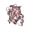
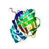
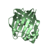
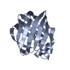
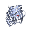
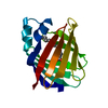
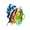
| Title | Crystal structure of human cellular retinol binding protein 3 in complex with abnormal-cannabidiol (abn-CBD) | ||||||
 Components Components | Retinol-binding protein 5 | ||||||
 Keywords Keywords | LIPID BINDING PROTEIN / vitamin A / retinol / abn-CBD / abnormal cannabidiol / cellular retinol-binding protein / CRBP3 | ||||||
| Function / homology |  Function and homology information Function and homology informationretinoid binding / retinal binding / retinol binding / fatty acid transport / fatty acid binding / extracellular exosome / nucleus / cytosol Similarity search - Function | ||||||
| Biological species |  Homo sapiens (human) Homo sapiens (human) | ||||||
| Method |  X-RAY DIFFRACTION / SYNCHROTRON / MOLECULAR REPLACEMENT / Resolution: 2.5 Å X-RAY DIFFRACTION / SYNCHROTRON / MOLECULAR REPLACEMENT / Resolution: 2.5 Å | ||||||
 Authors Authors | Silvaroli, J.A. / Banerjee, S. / Kiser, P.D. / Golczak, M. | ||||||
| Funding support |  United States, 1items United States, 1items
| ||||||
 Citation Citation | Journal: Acs Chem.Biol. / Year: 2019 Title: Abnormal Cannabidiol Modulates Vitamin A Metabolism by Acting as a Competitive Inhibitor of CRBP1. Authors: Silvaroli, J.A. / Widjaja-Adhi, M.A.K. / Trischman, T. / Chelstowska, S. / Horwitz, S. / Banerjee, S. / Kiser, P.D. / Blaner, W.S. / Golczak, M. | ||||||
| History |
|
- Structure visualization
| Structure viewer | Molecule: MolmilJmol/JSmol |
|---|
- Downloads & links
-Download
| PDBx/mmCIF format | 6e5w.cif.gz | 336.7 KB | Display | PDBx/mmCIF format |
|---|---|---|---|---|
| PDB format | pdb6e5w.ent.gz | 282.1 KB | Display | PDB format |
| PDBx/mmJSON format | 6e5w.json.gz | Tree view | PDBx/mmJSON format | |
| Others |  Other downloads Other downloads |
-Validation report
| Arichive directory | https://data.pdbj.org/pub/pdb/validation_reports/e5/6e5wftp://data.pdbj.org/pub/pdb/validation_reports/e5/6e5w | HTTPS FTP |
|---|
-Related structure data
| Related structure data | 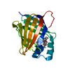 6e5lC 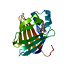 6e5tC  6e6kC 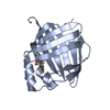 6e6mC 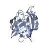 1gglS S: Starting model for refinement C: citing same article ( |
|---|---|
| Similar structure data |


-Links
 PDBj
PDBj

- Assembly
| Deposited unit | 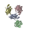
| ||||||||
|---|---|---|---|---|---|---|---|---|---|
| 1 | 
| ||||||||
| 2 | 
| ||||||||
| 3 | 
| ||||||||
| 4 | 
| ||||||||
| Unit cell |
|
-Components
| #1: Protein | Mass: 16419.840 Da / Num. of mol.: 4 Source method: isolated from a genetically manipulated source Source: (gene. exp.) Homo sapiens (human) / Gene: RBP5 / Production host:  #2: Chemical | ChemComp-HVD / (   Mass: 314.462 Da / Num. of mol.: 4 / Source method: obtained synthetically / Formula: C21H30O2 Mass: 314.462 Da / Num. of mol.: 4 / Source method: obtained synthetically / Formula: C21H30O2#3: Chemical | ChemComp-GOL /   Mass: 92.094 Da / Num. of mol.: 4 / Source method: isolated from a natural source / Formula: C3H8O3 Mass: 92.094 Da / Num. of mol.: 4 / Source method: isolated from a natural source / Formula: C3H8O3#4: Water | ChemComp-HOH / |  Mass: 18.015 Da / Num. of mol.: 208 / Source method: isolated from a natural source / Formula: H2O Mass: 18.015 Da / Num. of mol.: 208 / Source method: isolated from a natural source / Formula: H2O |
|---|
-Experimental details
-Experiment
| Experiment | Method: X-RAY DIFFRACTION / Number of used crystals: 1 |
|---|
- Sample preparation
| Crystal | Density Matthews: 2.91 Å3/Da / Density % sol: 57.78 % |
|---|---|
| Crystal grow | Temperature: 297 K / Method: vapor diffusion, sitting drop / pH: 5 / Details: 0.2M sodium malonate, pH 5.0 20% PEG 3350 |
-Data collection
| Diffraction | Mean temperature: 80 K |
|---|---|
| Diffraction source | Source: SYNCHROTRON / Site: APS / Beamline: 24-ID-C / Wavelength: 0.9791 Å |
| Detector | Type: DECTRIS PILATUS 6M / Detector: CCD / Date: Nov 5, 2017 |
| Radiation | Protocol: SINGLE WAVELENGTH / Monochromatic (M) / Laue (L): M / Scattering type: x-ray |
| Radiation wavelength | Wavelength: 0.9791 Å / Relative weight: 1 |
| Reflection | Resolution: 2.5→99.8 Å / Num. obs: 27540 / % possible obs: 99.9 % / Redundancy: 5.8 % / CC1/2: 0.99 / Rmerge(I) obs: 0.17 / Rpim(I) all: 0.08 / Net I/σ(I): 7.2 |
| Reflection shell | Resolution: 2.5→2.6 Å / Rmerge(I) obs: 0.87 / Mean I/σ(I) obs: 2.7 / Num. unique obs: 3005 / CC1/2: 0.8 / Rpim(I) all: 0.61 / % possible all: 99.9 |
- Processing
| Software |
| |||||||||||||||||||||||||||||||||||||||||||||||||||||||||||||||||||||||||||||
|---|---|---|---|---|---|---|---|---|---|---|---|---|---|---|---|---|---|---|---|---|---|---|---|---|---|---|---|---|---|---|---|---|---|---|---|---|---|---|---|---|---|---|---|---|---|---|---|---|---|---|---|---|---|---|---|---|---|---|---|---|---|---|---|---|---|---|---|---|---|---|---|---|---|---|---|---|---|---|
| Refinement | Method to determine structure: MOLECULAR REPLACEMENT Starting model: 1GGL Resolution: 2.5→99.773 Å / SU ML: 0.27 / Cross valid method: FREE R-VALUE / σ(F): 1.33 / Phase error: 26.64
| |||||||||||||||||||||||||||||||||||||||||||||||||||||||||||||||||||||||||||||
| Solvent computation | Shrinkage radii: 0.9 Å / VDW probe radii: 1.11 Å | |||||||||||||||||||||||||||||||||||||||||||||||||||||||||||||||||||||||||||||
| Refinement step | Cycle: LAST / Resolution: 2.5→99.773 Å
| |||||||||||||||||||||||||||||||||||||||||||||||||||||||||||||||||||||||||||||
| Refine LS restraints |
| |||||||||||||||||||||||||||||||||||||||||||||||||||||||||||||||||||||||||||||
| LS refinement shell |
| |||||||||||||||||||||||||||||||||||||||||||||||||||||||||||||||||||||||||||||
| Refinement TLS params. | Method: refined / Origin x: 37.6182 Å / Origin y: 0.2439 Å / Origin z: 18.3736 Å
| |||||||||||||||||||||||||||||||||||||||||||||||||||||||||||||||||||||||||||||
| Refinement TLS group | Selection details: all |
