 Movie
Movie Controller

[English] 日本語
 Yorodumi
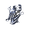
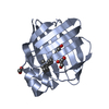
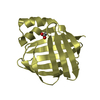
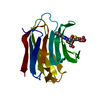
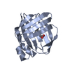
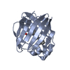
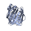
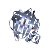
- PDB-1opb: THE CRYSTAL STRUCTURES OF HOLO-AND APO-CELLULAR RETINOL BINDING P... -
+ Open data

- Basic information
| Entry | Database: PDB / ID: 1opb | ||||||
|---|---|---|---|---|---|---|---|
| Title | THE CRYSTAL STRUCTURES OF HOLO-AND APO-CELLULAR RETINOL BINDING PROTEIN II | ||||||
 Components Components | CELLULAR RETINOL BINDING PROTEIN II | ||||||
 Keywords Keywords | RETINOL TRANSPORT | ||||||
| Function / homology |  Function and homology information Function and homology informationRetinoid metabolism and transport / triglyceride biosynthetic process / synaptic ribbon / retinal binding / retinol binding / retinol metabolic process / molecular carrier activity / fatty acid transport / retinoid metabolic process / fatty acid binding ...Retinoid metabolism and transport / triglyceride biosynthetic process / synaptic ribbon / retinal binding / retinol binding / retinol metabolic process / molecular carrier activity / fatty acid transport / retinoid metabolic process / fatty acid binding / transmembrane transporter binding / lipid binding / nucleus / cytosol Similarity search - Function | ||||||
| Biological species |  | ||||||
| Method |  X-RAY DIFFRACTION / Resolution: 1.9 Å X-RAY DIFFRACTION / Resolution: 1.9 Å | ||||||
 Authors Authors | Winter, N. / Banaszak, L. | ||||||
 Citation Citation | Journal: J.Mol.Biol. / Year: 1993 Title: Crystal structures of holo and apo-cellular retinol-binding protein II. Authors: Winter, N.S. / Bratt, J.M. / Banaszak, L.J. | ||||||
| History |
| ||||||
| Remark 700 | SHEET THE SHEET PRESENTED AS *S1* ON SHEET RECORDS BELOW IS ACTUALLY A TEN-STRANDED BETA-BARREL. ...SHEET THE SHEET PRESENTED AS *S1* ON SHEET RECORDS BELOW IS ACTUALLY A TEN-STRANDED BETA-BARREL. THIS IS REPRESENTED BY AN ELEVEN-STRANDED SHEET IN WHICH THE FIRST AND LAST STRANDS ARE IDENTICAL. |
- Structure visualization
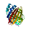
| Structure viewer | Molecule: MolmilJmol/JSmol |
|---|
- Downloads & links
-Download
| PDBx/mmCIF format | 1opb.cif.gz | 119.8 KB | Display | PDBx/mmCIF format |
|---|---|---|---|---|
| PDB format | pdb1opb.ent.gz | 95 KB | Display | PDB format |
| PDBx/mmJSON format | 1opb.json.gz | Tree view | PDBx/mmJSON format | |
| Others |  Other downloads Other downloads |
-Validation report
| Arichive directory | https://data.pdbj.org/pub/pdb/validation_reports/op/1opbftp://data.pdbj.org/pub/pdb/validation_reports/op/1opb | HTTPS FTP |
|---|
-Related structure data


-Links
 PDBj
PDBj


- Assembly
| Deposited unit | 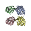
| ||||||||
|---|---|---|---|---|---|---|---|---|---|
| 1 | 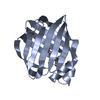
| ||||||||
| 2 | 
| ||||||||
| 3 | 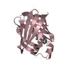
| ||||||||
| 4 | 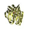
| ||||||||
| Unit cell |
|
-Components
| #1: Protein | Mass: 15606.570 Da / Num. of mol.: 4 Source method: isolated from a genetically manipulated source Source: (gene. exp.) #2: Chemical | ChemComp-RET /   Mass: 284.436 Da / Num. of mol.: 4 / Source method: obtained synthetically / Formula: C20H28O Mass: 284.436 Da / Num. of mol.: 4 / Source method: obtained synthetically / Formula: C20H28O#3: Water | ChemComp-HOH / |  Mass: 18.015 Da / Num. of mol.: 157 / Source method: isolated from a natural source / Formula: H2O Mass: 18.015 Da / Num. of mol.: 157 / Source method: isolated from a natural source / Formula: H2O |
|---|
-Experimental details
-Experiment
| Experiment | Method: X-RAY DIFFRACTION |
|---|
- Sample preparation
| Crystal | Density Matthews: 2.08 Å3/Da / Density % sol: 40.96 % | ||||||||||||||||||||||||||||||
|---|---|---|---|---|---|---|---|---|---|---|---|---|---|---|---|---|---|---|---|---|---|---|---|---|---|---|---|---|---|---|---|
| Crystal grow | *PLUS Temperature: 17.5 ℃ / pH: 4.5 / Method: vapor diffusion, hanging drop | ||||||||||||||||||||||||||||||
| Components of the solutions | *PLUS
|
-Data collection
| Reflection | *PLUS Highest resolution: 2.1 Å / Num. obs: 23764 / % possible obs: 75 % / Num. measured all: 57160 / Rmerge(I) obs: 0.0642 |
|---|
- Processing
| Software |
| ||||||||||||||||||||||||||||||||||||||||||||||||||||||||||||
|---|---|---|---|---|---|---|---|---|---|---|---|---|---|---|---|---|---|---|---|---|---|---|---|---|---|---|---|---|---|---|---|---|---|---|---|---|---|---|---|---|---|---|---|---|---|---|---|---|---|---|---|---|---|---|---|---|---|---|---|---|---|
| Refinement | Rfactor Rwork: 0.173 / Rfactor obs: 0.173 / Highest resolution: 1.9 Å Details: DATA WAS COLLECTED TO 2.1 ANGSTROMS RESOLUTION ON SIEMENS AREA DETECTOR. THE STRUCTURE WAS SOLVED BY MOLECULAR REPLACEMENT USING THE COORDINATES OF CELLULAR RETINOL BINDING PROTEIN AS A MODEL. | ||||||||||||||||||||||||||||||||||||||||||||||||||||||||||||
| Refinement step | Cycle: LAST / Highest resolution: 1.9 Å
| ||||||||||||||||||||||||||||||||||||||||||||||||||||||||||||
| Refine LS restraints |
| ||||||||||||||||||||||||||||||||||||||||||||||||||||||||||||
| Refinement | *PLUS Highest resolution: 2.1 Å / Rfactor obs: 0.173 / Num. reflection obs: 52976 / σ(F): 2 | ||||||||||||||||||||||||||||||||||||||||||||||||||||||||||||
| Solvent computation | *PLUS | ||||||||||||||||||||||||||||||||||||||||||||||||||||||||||||
| Displacement parameters | *PLUS Biso mean: 19.3 Å2 | ||||||||||||||||||||||||||||||||||||||||||||||||||||||||||||
| Refine LS restraints | *PLUS Type: x_angle_d / Dev ideal: 2.336 |
