 Movie
Movie Controller

[English] 日本語
 Yorodumi
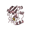
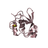
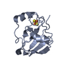
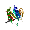
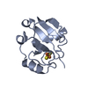
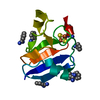
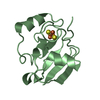
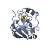
- PDB-1xlq: Crystal structure of reduced C73S putidaredoxin from Pseudomonas ... -
+ Open data

- Basic information
| Entry | Database: PDB / ID: 1xlq | ||||||
|---|---|---|---|---|---|---|---|
| Title | Crystal structure of reduced C73S putidaredoxin from Pseudomonas putida | ||||||
 Components Components | Putidaredoxin | ||||||
 Keywords Keywords | OXIDOREDUCTASE / [2Fe-2S] / ferredoxin | ||||||
| Function / homology |  Function and homology information Function and homology informationP450-containing electron transport chain / 2 iron, 2 sulfur cluster binding / electron transfer activity / metal ion binding / cytosol Similarity search - Function | ||||||
| Biological species |  Pseudomonas putida (bacteria) Pseudomonas putida (bacteria) | ||||||
| Method |  X-RAY DIFFRACTION / SYNCHROTRON / rigid body refinement / Resolution: 1.45 Å X-RAY DIFFRACTION / SYNCHROTRON / rigid body refinement / Resolution: 1.45 Å | ||||||
 Authors Authors | Sevrioukova, I.F. | ||||||
 Citation Citation | Journal: J.Mol.Biol. / Year: 2005 Title: Redox-dependent Structural Reorganization in Putidaredoxin, a Vertebrate-type [2Fe-2S] Ferredoxin from Pseudomonas putida. Authors: Sevrioukova, I.F. | ||||||
| History |
|
- Structure visualization
| Structure viewer | Molecule: MolmilJmol/JSmol |
|---|
- Downloads & links
-Download
| PDBx/mmCIF format | 1xlq.cif.gz | 79.6 KB | Display | PDBx/mmCIF format |
|---|---|---|---|---|
| PDB format | pdb1xlq.ent.gz | 59.6 KB | Display | PDB format |
| PDBx/mmJSON format | 1xlq.json.gz | Tree view | PDBx/mmJSON format | |
| Others |  Other downloads Other downloads |
-Validation report
| Arichive directory | https://data.pdbj.org/pub/pdb/validation_reports/xl/1xlqftp://data.pdbj.org/pub/pdb/validation_reports/xl/1xlq | HTTPS FTP |
|---|
-Related structure data
| Related structure data |  1xlnC  1xloC  1xlpC 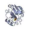 1oqrS S: Starting model for refinement C: citing same article ( |
|---|---|
| Similar structure data |


-Links
 PDBj
PDBj


- Assembly
| Deposited unit | 
| ||||||||
|---|---|---|---|---|---|---|---|---|---|
| 1 | 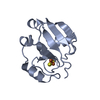
| ||||||||
| 2 | 
| ||||||||
| 3 | 
| ||||||||
| 4 | 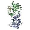
| ||||||||
| Unit cell |
|
-Components
| #1: Protein | Mass: 11412.846 Da / Num. of mol.: 3 / Mutation: C73S Source method: isolated from a genetically manipulated source Source: (gene. exp.) Pseudomonas putida (bacteria) / Gene: camB / Plasmid: pET / Species (production host): Escherichia coli / Production host: #2: Chemical |   Mass: 175.820 Da / Num. of mol.: 3 / Source method: obtained synthetically / Formula: Fe2S2 Mass: 175.820 Da / Num. of mol.: 3 / Source method: obtained synthetically / Formula: Fe2S2#3: Water | ChemComp-HOH / |  Mass: 18.015 Da / Num. of mol.: 349 / Source method: isolated from a natural source / Formula: H2O Mass: 18.015 Da / Num. of mol.: 349 / Source method: isolated from a natural source / Formula: H2O |
|---|
-Experimental details
-Experiment
| Experiment | Method: X-RAY DIFFRACTION / Number of used crystals: 1 |
|---|
- Sample preparation
| Crystal | Density Matthews: 2.3 Å3/Da / Density % sol: 45.5 % |
|---|---|
| Crystal grow | Temperature: 293 K / Method: vapor diffusion, hanging drop / pH: 5.7 Details: lithium sulfate, sodium citrate, pH 5.7, VAPOR DIFFUSION, HANGING DROP, temperature 293K |
-Data collection
| Diffraction | Mean temperature: 170 K |
|---|---|
| Diffraction source | Source: SYNCHROTRON / Site: SSRL  / Beamline: BL7-1 / Wavelength: 1.08 Å / Beamline: BL7-1 / Wavelength: 1.08 Å |
| Detector | Type: MARRESEARCH / Detector: IMAGE PLATE / Date: Dec 18, 2002 / Details: mirrors |
| Radiation | Monochromator: Yale mirrors / Protocol: SINGLE WAVELENGTH / Monochromatic (M) / Laue (L): M / Scattering type: x-ray |
| Radiation wavelength | Wavelength: 1.08 Å / Relative weight: 1 |
| Reflection | Resolution: 1.45→27.92 Å / Num. all: 61499 / Num. obs: 60208 / % possible obs: 97.9 % / Observed criterion σ(I): -3 / Redundancy: 3.9 % / Biso Wilson estimate: 16.5 Å2 / Rmerge(I) obs: 0.044 / Rsym value: 0.044 / Net I/σ(I): 28.8 |
| Reflection shell | Resolution: 1.45→1.5 Å / Redundancy: 3.5 % / Rmerge(I) obs: 0.545 / Mean I/σ(I) obs: 2.6 / Num. unique all: 5335 / Rsym value: 0.545 / % possible all: 92.7 |
- Processing
| Software |
| |||||||||||||||||||||||||
|---|---|---|---|---|---|---|---|---|---|---|---|---|---|---|---|---|---|---|---|---|---|---|---|---|---|---|
| Refinement | Method to determine structure: rigid body refinement Starting model: 1oqr Resolution: 1.45→27.92 Å / Rfactor Rfree error: 0.004 / Isotropic thermal model: isotropic / Cross valid method: THROUGHOUT / σ(I): -3 / Stereochemistry target values: Engh & Huber
| |||||||||||||||||||||||||
| Solvent computation | Solvent model: Flat Model / Bsol: 42.6 Å2 / ksol: 0.37 e/Å3 | |||||||||||||||||||||||||
| Displacement parameters | Biso mean: 21 Å2 | |||||||||||||||||||||||||
| Refine analyze |
| |||||||||||||||||||||||||
| Refinement step | Cycle: LAST / Resolution: 1.45→27.92 Å
| |||||||||||||||||||||||||
| Refine LS restraints |
| |||||||||||||||||||||||||
| LS refinement shell | Resolution: 1.45→1.46 Å / Rfactor Rfree error: 0.004 / Total num. of bins used: 10
|
