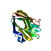
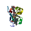
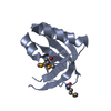
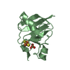
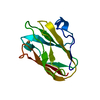
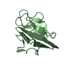
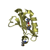
- PDB-3plu: Structure of Hub-1 protein in complex with Snu66 peptide (HINDI) -
+
Open data
ID or keywords:
Loading...
-
Basic information
Entry
Database: PDB / ID: 3plu
Title
Structure of Hub-1 protein in complex with Snu66 peptide (HINDI)
Components
66 kDa U4/U6.U5 small nuclear ribonucleoprotein component
Ubiquitin-like modifier HUB1
Keywords
PEPTIDE BINDING PROTEIN / Ubiquitin-like / HUB-1 / Snu66
Function / homology
Function and homology information
cell morphogenesis involved in conjugation with cellular fusion / maturation of 5S rRNA / mRNA cis splicing, via spliceosome / U4/U6 x U5 tri-snRNP complex / post-translational protein modification / positive regulation of RNA splicing / spliceosomal complex / mRNA splicing, via spliceosome / protein tag activity / nucleus / cytoplasm Similarity search - Function
Ubiquitin-like modifier Hub1/Ubl5 / SNU66/SART1 family / HIND motif / SART-1 family / HIND motif / Phosphatidylinositol 3-kinase Catalytic Subunit; Chain A, domain 1 / Ubiquitin-like (UB roll) / Ubiquitin family / Ubiquitin domain profile. / Ubiquitin-like domain ...Ubiquitin-like modifier Hub1/Ubl5 / SNU66/SART1 family / HIND motif / SART-1 family / HIND motif / Phosphatidylinositol 3-kinase Catalytic Subunit; Chain A, domain 1 / Ubiquitin-like (UB roll) / Ubiquitin family / Ubiquitin domain profile. / Ubiquitin-like domain / Ubiquitin-like domain superfamily / Roll / Alpha Beta Similarity search - Domain/homology
Resolution: 1.4→26.12 Å / Cor.coef. Fo:Fc: 0.963 / Cor.coef. Fo:Fc free: 0.941 / SU B: 2.433 / SU ML: 0.045 / Cross valid method: THROUGHOUT / σ(F): 2 / ESU R Free: 0.077 / Stereochemistry target values: MAXIMUM LIKELIHOOD / Details: HYDROGENS HAVE BEEN ADDED IN THE RIDING POSITIONS
Rfactor
Num. reflection
% reflection
Selection details
Rfree
0.22819
1686
5 %
RANDOM
Rwork
0.18521
-
-
-
obs
0.18737
31992
94.63 %
-
all
-
33677
-
-
Solvent computation
Ion probe radii: 0.8 Å / Shrinkage radii: 0.8 Å / VDW probe radii: 1.4 Å / Solvent model: MASK
Displacement parameters
Biso mean: 18.696 Å2
Baniso -1
Baniso -2
Baniso -3
1-
-0.12 Å2
0.11 Å2
-0.05 Å2
2-
-
0.17 Å2
-0.01 Å2
3-
-
-
-0.07 Å2
Refinement step
Cycle: LAST / Resolution: 1.4→26.12 Å
Protein
Nucleic acid
Ligand
Solvent
Total
Num. atoms
1443
0
0
275
1718
Refine LS restraints
Refine-ID
Type
Dev ideal
Dev ideal target
Number
X-RAY DIFFRACTION
r_bond_refined_d
0.007
0.022
1457
X-RAY DIFFRACTION
r_angle_refined_deg
1.033
1.997
1963
X-RAY DIFFRACTION
r_dihedral_angle_1_deg
5.229
5
180
X-RAY DIFFRACTION
r_dihedral_angle_2_deg
38.795
26
65
X-RAY DIFFRACTION
r_dihedral_angle_3_deg
10.527
15
284
X-RAY DIFFRACTION
r_dihedral_angle_4_deg
7.488
15
6
X-RAY DIFFRACTION
r_chiral_restr
0.073
0.2
234
X-RAY DIFFRACTION
r_gen_planes_refined
0.003
0.02
1056
X-RAY DIFFRACTION
r_nbd_refined
0.177
0.2
668
X-RAY DIFFRACTION
r_nbtor_refined
0.298
0.2
986
X-RAY DIFFRACTION
r_xyhbond_nbd_refined
0.096
0.2
203
X-RAY DIFFRACTION
r_symmetry_vdw_refined
0.185
0.2
39
X-RAY DIFFRACTION
r_symmetry_hbond_refined
0.1
0.2
30
X-RAY DIFFRACTION
r_mcbond_it
1.125
1.5
939
X-RAY DIFFRACTION
r_mcangle_it
1.704
2
1471
X-RAY DIFFRACTION
r_scbond_it
1.885
3
569
X-RAY DIFFRACTION
r_scangle_it
2.934
4.5
492
X-RAY DIFFRACTION
r_rigid_bond_restr
1.13
3
1508
X-RAY DIFFRACTION
r_sphericity_free
2.956
3
276
X-RAY DIFFRACTION
r_sphericity_bonded
3.273
3
1443
LS refinement shell
Resolution: 1.4→1.436 Å / Total num. of bins used: 20
Rfactor
Num. reflection
% reflection
Rfree
0.296
119
-
Rwork
0.242
2268
-
obs
-
-
91.98 %
+
About Yorodumi
-
News
-
Feb 9, 2022. New format data for meta-information of EMDB entries
New format data for meta-information of EMDB entries
Version 3 of the EMDB header file is now the official format.
The previous official version 1.9 will be removed from the archive.
In the structure databanks used in Yorodumi, some data are registered as the other names, "COVID-19 virus" and "2019-nCoV". Here are the details of the virus and the list of structure data.
Jan 31, 2019. EMDB accession codes are about to change! (news from PDBe EMDB page)
EMDB accession codes are about to change! (news from PDBe EMDB page)
The allocation of 4 digits for EMDB accession codes will soon come to an end. Whilst these codes will remain in use, new EMDB accession codes will include an additional digit and will expand incrementally as the available range of codes is exhausted. The current 4-digit format prefixed with “EMD-” (i.e. EMD-XXXX) will advance to a 5-digit format (i.e. EMD-XXXXX), and so on. It is currently estimated that the 4-digit codes will be depleted around Spring 2019, at which point the 5-digit format will come into force.
The EM Navigator/Yorodumi systems omit the EMD- prefix.
Related info.:Q: What is EMD? / ID/Accession-code notation in Yorodumi/EM Navigator
Yorodumi is a browser for structure data from EMDB, PDB, SASBDB, etc.
This page is also the successor to EM Navigator detail page, and also detail information page/front-end page for Omokage search.
The word "yorodu" (or yorozu) is an old Japanese word meaning "ten thousand". "mi" (miru) is to see.
Related info.:EMDB / PDB / SASBDB / Comparison of 3 databanks / Yorodumi Search / Aug 31, 2016. New EM Navigator & Yorodumi / Yorodumi Papers / Jmol/JSmol / Function and homology information / Changes in new EM Navigator and Yorodumi
 Movie
Movie Controller


 Open data
 Basic information
 Components
 Keywords
 Function and homology information
 X-RAY DIFFRACTION /
  Authors
 Citation
 Structure visualization
 Downloads & links
 Other downloads


 PDBj
PDBj Assembly


 Mass: 18.015 Da / Num. of mol.: 275 / Source method: isolated from a natural source / Formula: H2O
 Sample preparation
 / Beamline: BW6 / Wavelength: 1.05 Å
 Processing