 Movie
Movie Controller

[English] 日本語
 Yorodumi
- PDB-3av8: Refined Structure of Plant-type [2Fe-2S] Ferredoxin I from Aphano... -
+ Open data

- Basic information
| Entry | Database: PDB / ID: 3av8 | ||||||
|---|---|---|---|---|---|---|---|
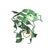
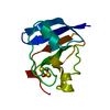
| Title | Refined Structure of Plant-type [2Fe-2S] Ferredoxin I from Aphanothece sacrum at 1.46 A Resolution | ||||||
 Components Components | Ferredoxin-1 | ||||||
 Keywords Keywords | ELECTRON TRANSPORT / Beta-grasp / Redox Protein | ||||||
| Function / homology |  Function and homology information Function and homology informationelectron transport chain / 2 iron, 2 sulfur cluster binding / electron transfer activity / metal ion binding Similarity search - Function | ||||||
| Biological species |  Aphanothece sacrum (bacteria) Aphanothece sacrum (bacteria) | ||||||
| Method |  X-RAY DIFFRACTION / SYNCHROTRON / MOLECULAR REPLACEMENT / Resolution: 1.46 Å X-RAY DIFFRACTION / SYNCHROTRON / MOLECULAR REPLACEMENT / Resolution: 1.46 Å | ||||||
 Authors Authors | Kameda, H. / Hirabayashi, K. / Wada, K. / Fukuyama, K. | ||||||
 Citation Citation | Journal: Plos One / Year: 2011 Title: Mapping of protein-protein interaction sites in the plant-type [2Fe-2S] ferredoxin. Authors: Kameda, H. / Hirabayashi, K. / Wada, K. / Fukuyama, K. | ||||||
| History |
|
- Structure visualization
| Structure viewer | Molecule: MolmilJmol/JSmol |
|---|
- Downloads & links
-Download
| PDBx/mmCIF format | 3av8.cif.gz | 96 KB | Display | PDBx/mmCIF format |
|---|---|---|---|---|
| PDB format | pdb3av8.ent.gz | 74.1 KB | Display | PDB format |
| PDBx/mmJSON format | 3av8.json.gz | Tree view | PDBx/mmJSON format | |
| Others |  Other downloads Other downloads |
-Validation report
| Arichive directory | https://data.pdbj.org/pub/pdb/validation_reports/av/3av8ftp://data.pdbj.org/pub/pdb/validation_reports/av/3av8 | HTTPS FTP |
|---|
-Related structure data
| Related structure data |  1fxiS S: Starting model for refinement |
|---|---|
| Similar structure data |


-Links
 PDBj
PDBj


- Assembly
| Deposited unit | 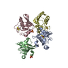
| ||||||||
|---|---|---|---|---|---|---|---|---|---|
| 1 | 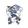
| ||||||||
| 2 | 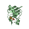
| ||||||||
| 3 | 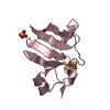
| ||||||||
| 4 | 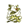
| ||||||||
| Unit cell |
|
-Components
| #1: Protein | Mass: 10399.398 Da / Num. of mol.: 4 Source method: isolated from a genetically manipulated source Source: (gene. exp.) Aphanothece sacrum (bacteria) / Plasmid: pET21a / Production host: #2: Chemical | ChemComp-FES /   Mass: 175.820 Da / Num. of mol.: 4 / Source method: obtained synthetically / Formula: Fe2S2 Mass: 175.820 Da / Num. of mol.: 4 / Source method: obtained synthetically / Formula: Fe2S2#3: Chemical |   Mass: 96.063 Da / Num. of mol.: 2 / Source method: obtained synthetically / Formula: SO4 Mass: 96.063 Da / Num. of mol.: 2 / Source method: obtained synthetically / Formula: SO4#4: Water | ChemComp-HOH / |  Mass: 18.015 Da / Num. of mol.: 485 / Source method: isolated from a natural source / Formula: H2O Mass: 18.015 Da / Num. of mol.: 485 / Source method: isolated from a natural source / Formula: H2O |
|---|
-Experimental details
-Experiment
| Experiment | Method: X-RAY DIFFRACTION / Number of used crystals: 1 |
|---|
- Sample preparation
| Crystal | Density Matthews: 2.36 Å3/Da / Density % sol: 47.79 % |
|---|---|
| Crystal grow | Temperature: 277 K / Method: microdialysis / pH: 7.5 Details: 70-80% ammonium sulfate, 0.1M TRIS, 0.7M sodium chloride, pH 7.5, MICRODIALYSIS, temperature 277K |
-Data collection
| Diffraction | Mean temperature: 100 K |
|---|---|
| Diffraction source | Source: SYNCHROTRON / Site: SPring-8  / Beamline: BL32XU / Wavelength: 1 Å / Beamline: BL32XU / Wavelength: 1 Å |
| Detector | Type: RAYONIX MX225HE / Detector: CCD / Date: Dec 15, 2010 |
| Radiation | Monochromator: A Double Crystal Monochromator / Protocol: SINGLE WAVELENGTH / Monochromatic (M) / Laue (L): M / Scattering type: x-ray |
| Radiation wavelength | Wavelength: 1 Å / Relative weight: 1 |
| Reflection | Resolution: 1.46→25 Å / Num. obs: 66810 / % possible obs: 99 % / Observed criterion σ(F): 0 / Observed criterion σ(I): 0 / Redundancy: 5.7 % / Biso Wilson estimate: 20.2 Å2 / Rsym value: 0.05 |
| Reflection shell | Resolution: 1.46→1.51 Å / Redundancy: 5.5 % / Rsym value: 0.383 / % possible all: 99.6 |
- Processing
| Software |
| ||||||||||||||||||||||||||||||||||||
|---|---|---|---|---|---|---|---|---|---|---|---|---|---|---|---|---|---|---|---|---|---|---|---|---|---|---|---|---|---|---|---|---|---|---|---|---|---|
| Refinement | Method to determine structure: MOLECULAR REPLACEMENT Starting model: PDB ENTRY 1FXI Resolution: 1.46→24.64 Å / Rfactor Rfree error: 0.004 / Data cutoff high absF: 269412.93 / Data cutoff low absF: 0 / Isotropic thermal model: RESTRAINED / Cross valid method: THROUGHOUT / σ(F): 0 / Stereochemistry target values: Engh & Huber
| ||||||||||||||||||||||||||||||||||||
| Solvent computation | Solvent model: FLAT MODEL / Bsol: 61.1651 Å2 / ksol: 0.405467 e/Å3 | ||||||||||||||||||||||||||||||||||||
| Displacement parameters | Biso mean: 28 Å2
| ||||||||||||||||||||||||||||||||||||
| Refine analyze |
| ||||||||||||||||||||||||||||||||||||
| Refinement step | Cycle: LAST / Resolution: 1.46→24.64 Å
| ||||||||||||||||||||||||||||||||||||
| Refine LS restraints |
| ||||||||||||||||||||||||||||||||||||
| LS refinement shell | Resolution: 1.46→1.55 Å / Rfactor Rfree error: 0.013 / Total num. of bins used: 6
| ||||||||||||||||||||||||||||||||||||
| Xplor file |
|
