 Movie
Movie Controller


+ Open data

- Basic information
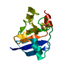
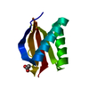
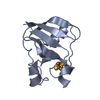
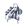
| Entry | Database: PDB / ID: 1a70 | ||||||
|---|---|---|---|---|---|---|---|
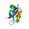
| Title | SPINACH FERREDOXIN | ||||||
 Components Components | FERREDOXIN | ||||||
 Keywords Keywords | IRON-SULFUR PROTEIN / PHOTOSYNTHESIS / ELECTRON TRANSPORT | ||||||
| Function / homology |  Function and homology information Function and homology informationchloroplast stroma / electron transport chain / 2 iron, 2 sulfur cluster binding / electron transfer activity / metal ion binding Similarity search - Function | ||||||
| Biological species |  Spinacia oleracea (spinach) Spinacia oleracea (spinach) | ||||||
| Method |  X-RAY DIFFRACTION / MOLECULAR REPLACEMENT / Resolution: 1.7 Å X-RAY DIFFRACTION / MOLECULAR REPLACEMENT / Resolution: 1.7 Å | ||||||
 Authors Authors | Binda, C. / Coda, A. / Mattevi, A. / Aliverti, A. / Zanetti, G. | ||||||
 Citation Citation | Journal: Acta Crystallogr.,Sect.D / Year: 1998 Title: Structure of the mutant E92K of [2Fe-2S] ferredoxin I from Spinacia oleracea at 1.7 A resolution. Authors: Binda, C. / Coda, A. / Aliverti, A. / Zanetti, G. / Mattevi, A. | ||||||
| History |
|
- Structure visualization
| Structure viewer | Molecule: MolmilJmol/JSmol |
|---|
- Downloads & links
-Download
| PDBx/mmCIF format | 1a70.cif.gz | 32.8 KB | Display | PDBx/mmCIF format |
|---|---|---|---|---|
| PDB format | pdb1a70.ent.gz | 20.9 KB | Display | PDB format |
| PDBx/mmJSON format | 1a70.json.gz | Tree view | PDBx/mmJSON format | |
| Others |  Other downloads Other downloads |
-Validation report
| Arichive directory | https://data.pdbj.org/pub/pdb/validation_reports/a7/1a70ftp://data.pdbj.org/pub/pdb/validation_reports/a7/1a70 | HTTPS FTP |
|---|
-Related structure data
| Related structure data |  1frrS S: Starting model for refinement |
|---|---|
| Similar structure data |


-Links
 PDBj
PDBj


- Assembly
| Deposited unit | 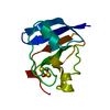
| ||||||||
|---|---|---|---|---|---|---|---|---|---|
| 1 |
| ||||||||
| Unit cell |
|
-Components
| #1: Protein | Mass: 10488.470 Da / Num. of mol.: 1 / Mutation: E92K Source method: isolated from a genetically manipulated source Source: (gene. exp.) Spinacia oleracea (spinach) / Production host:  |
|---|---|
| #2: Chemical | ChemComp-FES /   Mass: 175.820 Da / Num. of mol.: 1 / Source method: obtained synthetically / Formula: Fe2S2 Mass: 175.820 Da / Num. of mol.: 1 / Source method: obtained synthetically / Formula: Fe2S2 |
| #3: Water | ChemComp-HOH /  Mass: 18.015 Da / Num. of mol.: 81 / Source method: isolated from a natural source / Formula: H2O Mass: 18.015 Da / Num. of mol.: 81 / Source method: isolated from a natural source / Formula: H2O |
-Experimental details
-Experiment
| Experiment | Method: X-RAY DIFFRACTION / Number of used crystals: 1 |
|---|
- Sample preparation
| Crystal | Density Matthews: 1.5 Å3/Da / Density % sol: 50 % | ||||||||||||||||||||||||||||||
|---|---|---|---|---|---|---|---|---|---|---|---|---|---|---|---|---|---|---|---|---|---|---|---|---|---|---|---|---|---|---|---|
| Crystal grow | pH: 7.5 Details: PROTEIN WAS CRYSTALLIZED FROM 2.6M AMMONIUM SULPHATE IN 50MM PHOSPHATE BUFFER, PH 7.5 | ||||||||||||||||||||||||||||||
| Crystal grow | *PLUS Temperature: 277 K / Method: vapor diffusion, hanging drop | ||||||||||||||||||||||||||||||
| Components of the solutions | *PLUS
|
-Data collection
| Diffraction | Mean temperature: 293 K |
|---|---|
| Diffraction source | Source: ROTATING ANODE / Type: RIGAKU RUH2R / Wavelength: 1.5418 |
| Detector | Type: MARRESEARCH / Detector: IMAGE PLATE / Date: Jun 1, 1996 / Details: MIRRORS |
| Radiation | Monochromator: GRAPHITE(002) / Monochromatic (M) / Laue (L): M / Scattering type: x-ray |
| Radiation wavelength | Wavelength: 1.5418 Å / Relative weight: 1 |
| Reflection | Highest resolution: 1.7 Å / Num. obs: 11755 / % possible obs: 97.6 % / Observed criterion σ(I): 2 / Redundancy: 2.5 % / Biso Wilson estimate: 5.1 Å2 / Rmerge(I) obs: 0.078 / Rsym value: 0.112 / Net I/σ(I): 10 |
| Reflection shell | Resolution: 1.7→1.8 Å / Redundancy: 2.5 % / Rmerge(I) obs: 0.202 / Mean I/σ(I) obs: 2 / Rsym value: 0.305 / % possible all: 68 |
| Reflection | *PLUS Num. measured all: 35600 |
| Reflection shell | *PLUS % possible obs: 68 % |
- Processing
| Software |
| ||||||||||||||||||||||||||||||||||||||||||||||||||
|---|---|---|---|---|---|---|---|---|---|---|---|---|---|---|---|---|---|---|---|---|---|---|---|---|---|---|---|---|---|---|---|---|---|---|---|---|---|---|---|---|---|---|---|---|---|---|---|---|---|---|---|
| Refinement | Method to determine structure: MOLECULAR REPLACEMENT Starting model: PDB ENTRY 1FRR Resolution: 1.7→100 Å / Isotropic thermal model: TNT BCORREL V1.0 / Cross valid method: A POSTERIORI / σ(F): 0 / Stereochemistry target values: TNT PROTGEO
| ||||||||||||||||||||||||||||||||||||||||||||||||||
| Solvent computation | Solvent model: BABINET SCALING / Bsol: 150 Å2 / ksol: 0.8 e/Å3 | ||||||||||||||||||||||||||||||||||||||||||||||||||
| Refinement step | Cycle: LAST / Resolution: 1.7→100 Å
| ||||||||||||||||||||||||||||||||||||||||||||||||||
| Refine LS restraints |
| ||||||||||||||||||||||||||||||||||||||||||||||||||
| Software | *PLUS Name: TNT / Version: 5D / Classification: refinement | ||||||||||||||||||||||||||||||||||||||||||||||||||
| Refinement | *PLUS | ||||||||||||||||||||||||||||||||||||||||||||||||||
| Solvent computation | *PLUS | ||||||||||||||||||||||||||||||||||||||||||||||||||
| Displacement parameters | *PLUS | ||||||||||||||||||||||||||||||||||||||||||||||||||
| Refine LS restraints | *PLUS
|
