 Movie
Movie Controller

[English] 日本語
 Yorodumi
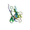
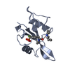
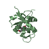
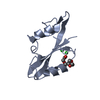
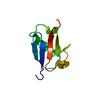
- PDB-3plv: Structure of Hub-1 protein in complex with Snu66 peptide (HINDII) -
+ Open data

- Basic information
| Entry | Database: PDB / ID: 3plv | ||||||
|---|---|---|---|---|---|---|---|
| Title | Structure of Hub-1 protein in complex with Snu66 peptide (HINDII) | ||||||
 Components Components |
| ||||||
 Keywords Keywords | PEPTIDE BINDING PROTEIN / Ubiquitin-like | ||||||
| Function / homology |  Function and homology information Function and homology informationcell morphogenesis involved in conjugation with cellular fusion / maturation of 5S rRNA / mRNA cis splicing, via spliceosome / U4/U6 x U5 tri-snRNP complex / post-translational protein modification / positive regulation of RNA splicing / spliceosomal complex / mRNA splicing, via spliceosome / protein tag activity / nucleus / cytoplasm Similarity search - Function | ||||||
| Biological species |  | ||||||
| Method |  X-RAY DIFFRACTION / SYNCHROTRON / MOLECULAR REPLACEMENT / Resolution: 1.9 Å X-RAY DIFFRACTION / SYNCHROTRON / MOLECULAR REPLACEMENT / Resolution: 1.9 Å | ||||||
 Authors Authors | Mishra, S.K. / Ammon, T. / Popowicz, G.M. / Krajewski, M. / Nagel, R.J. / Ares, M. / Holak, T.A. / Jentsch, S. | ||||||
 Citation Citation | Journal: Nature / Year: 2011 Title: Role of the ubiquitin-like protein Hub1 in splice-site usage and alternative splicing. Authors: Mishra, S.K. / Ammon, T. / Popowicz, G.M. / Krajewski, M. / Nagel, R.J. / Ares, M. / Holak, T.A. / Jentsch, S. | ||||||
| History |
|
- Structure visualization
| Structure viewer | Molecule: MolmilJmol/JSmol |
|---|
- Downloads & links
-Download
| PDBx/mmCIF format | 3plv.cif.gz | 36.2 KB | Display | PDBx/mmCIF format |
|---|---|---|---|---|
| PDB format | pdb3plv.ent.gz | 23.7 KB | Display | PDB format |
| PDBx/mmJSON format | 3plv.json.gz | Tree view | PDBx/mmJSON format | |
| Others |  Other downloads Other downloads |
-Validation report
| Arichive directory | https://data.pdbj.org/pub/pdb/validation_reports/pl/3plvftp://data.pdbj.org/pub/pdb/validation_reports/pl/3plv | HTTPS FTP |
|---|
-Related structure data
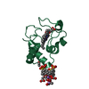
| Related structure data |  3pluC  1ubiS C: citing same article ( S: Starting model for refinement |
|---|---|
| Similar structure data |


-Links
 PDBj
PDBj- Assembly
| Deposited unit | 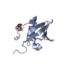
| ||||||||
|---|---|---|---|---|---|---|---|---|---|
| 1 |
| ||||||||
| Unit cell |
|
-Components
| #1: Protein | Mass: 10440.913 Da / Num. of mol.: 1 / Mutation: A20G Source method: isolated from a genetically manipulated source Source: (gene. exp.) Gene: HUB1, YNR032C-A / Plasmid: pET-28 / Production host:  |
|---|---|
| #2: Protein/peptide | Mass: 2282.634 Da / Num. of mol.: 1 / Fragment: HINDII domain / Source method: obtained synthetically / Source: (synth.) |
| #3: Water | ChemComp-HOH /  Mass: 18.015 Da / Num. of mol.: 157 / Source method: isolated from a natural source / Formula: H2O Mass: 18.015 Da / Num. of mol.: 157 / Source method: isolated from a natural source / Formula: H2O |
-Experimental details
-Experiment
| Experiment | Method: X-RAY DIFFRACTION / Number of used crystals: 1 |
|---|
- Sample preparation
| Crystal | Density Matthews: 2.12 Å3/Da / Density % sol: 41.9 % |
|---|---|
| Crystal grow | Temperature: 290 K / Method: vapor diffusion, sitting drop / pH: 6.9 Details: 0.2M Ammonium Iodide, 20% PEG 3350, pH 6.9, VAPOR DIFFUSION, SITTING DROP, temperature 290K |
-Data collection
| Diffraction | Mean temperature: 90 K |
|---|---|
| Diffraction source | Source: SYNCHROTRON / Site: MPG/DESY, HAMBURG  / Beamline: BW6 / Wavelength: 1.05 Å / Beamline: BW6 / Wavelength: 1.05 Å |
| Detector | Type: MAR CCD 165 mm / Detector: CCD / Date: May 9, 2006 / Details: mirrors |
| Radiation | Monochromator: Si-111 / Protocol: SINGLE WAVELENGTH / Monochromatic (M) / Laue (L): M / Scattering type: x-ray |
| Radiation wavelength | Wavelength: 1.05 Å / Relative weight: 1 |
| Reflection | Resolution: 1.9→45 Å / Num. all: 9009 / Num. obs: 8959 / % possible obs: 99.4 % / Observed criterion σ(F): 2 / Observed criterion σ(I): 2 |
| Reflection shell | Resolution: 1.9→2 Å / Rmerge(I) obs: 0.136 / Mean I/σ(I) obs: 9.96 / % possible all: 97.8 |
- Processing
| Software |
| ||||||||||||||||||||||||||||||||||||||||||||||||||||||||||||||||||||||||||||||||||||||||||||||||||||||||||||||||||||||||||||||||||||||||||||||||||||||||||||||||||||||||||
|---|---|---|---|---|---|---|---|---|---|---|---|---|---|---|---|---|---|---|---|---|---|---|---|---|---|---|---|---|---|---|---|---|---|---|---|---|---|---|---|---|---|---|---|---|---|---|---|---|---|---|---|---|---|---|---|---|---|---|---|---|---|---|---|---|---|---|---|---|---|---|---|---|---|---|---|---|---|---|---|---|---|---|---|---|---|---|---|---|---|---|---|---|---|---|---|---|---|---|---|---|---|---|---|---|---|---|---|---|---|---|---|---|---|---|---|---|---|---|---|---|---|---|---|---|---|---|---|---|---|---|---|---|---|---|---|---|---|---|---|---|---|---|---|---|---|---|---|---|---|---|---|---|---|---|---|---|---|---|---|---|---|---|---|---|---|---|---|---|---|---|---|
| Refinement | Method to determine structure: MOLECULAR REPLACEMENT Starting model: PDB entry 1UBI Resolution: 1.9→41.78 Å / Cor.coef. Fo:Fc: 0.925 / Cor.coef. Fo:Fc free: 0.891 / SU B: 3.91 / SU ML: 0.116 / Cross valid method: THROUGHOUT / σ(F): 2 / ESU R Free: 0.155 / Stereochemistry target values: MAXIMUM LIKELIHOOD / Details: HYDROGENS HAVE BEEN ADDED IN THE RIDING POSITIONS
| ||||||||||||||||||||||||||||||||||||||||||||||||||||||||||||||||||||||||||||||||||||||||||||||||||||||||||||||||||||||||||||||||||||||||||||||||||||||||||||||||||||||||||
| Solvent computation | Ion probe radii: 0.8 Å / Shrinkage radii: 0.8 Å / VDW probe radii: 1.4 Å / Solvent model: MASK | ||||||||||||||||||||||||||||||||||||||||||||||||||||||||||||||||||||||||||||||||||||||||||||||||||||||||||||||||||||||||||||||||||||||||||||||||||||||||||||||||||||||||||
| Displacement parameters | Biso mean: 11.774 Å2
| ||||||||||||||||||||||||||||||||||||||||||||||||||||||||||||||||||||||||||||||||||||||||||||||||||||||||||||||||||||||||||||||||||||||||||||||||||||||||||||||||||||||||||
| Refinement step | Cycle: LAST / Resolution: 1.9→41.78 Å
| ||||||||||||||||||||||||||||||||||||||||||||||||||||||||||||||||||||||||||||||||||||||||||||||||||||||||||||||||||||||||||||||||||||||||||||||||||||||||||||||||||||||||||
| Refine LS restraints |
| ||||||||||||||||||||||||||||||||||||||||||||||||||||||||||||||||||||||||||||||||||||||||||||||||||||||||||||||||||||||||||||||||||||||||||||||||||||||||||||||||||||||||||
| LS refinement shell | Resolution: 1.9→1.949 Å / Total num. of bins used: 20
|
