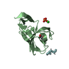
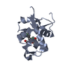
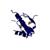
Mass: 10861.375 Da / Num. of mol.: 1 Source method: isolated from a genetically manipulated source Source: (gene. exp.) Vibrio cholerae serotype O1 (bacteria) / Strain: ATCC 39315 / El Tor Inaba N16961 / Gene: VC_1496 / Plasmid: pMCSG53 / Production host: Escherichia coli BL21(DE3) (bacteria) / Strain (production host): BL21 (DE3) magic / References: UniProt: Q9KRY7
#2: Protein/peptide
LEU-ILE-ALA
Mass: 315.409 Da / Num. of mol.: 1 / Source method: isolated from a natural source Details: peptide belongs to not identified protein from E. coli. Source: (natural) Escherichia coli (E. coli)
Method to determine structure: SAD / Resolution: 1.45→29.82 Å / Cor.coef. Fo:Fc: 0.982 / Cor.coef. Fo:Fc free: 0.969 / SU B: 3.1 / SU ML: 0.052 / Cross valid method: THROUGHOUT / ESU R: 0.074 / ESU R Free: 0.066 / Details: HYDROGENS HAVE BEEN ADDED IN THE RIDING POSITIONS
Rfactor
Num. reflection
% reflection
Selection details
Rfree
0.17758
708
4.7 %
RANDOM
Rwork
0.1324
-
-
-
obs
0.1346
14288
99.94 %
-
Solvent computation
Ion probe radii: 0.8 Å / Shrinkage radii: 0.8 Å / VDW probe radii: 1.2 Å
 Movie
Movie Controller

 Yorodumi
 Open data
 Basic information
 Components
 Keywords
 Function and homology information
 Vibrio cholerae serotype O1 (bacteria)
 X-RAY DIFFRACTION /
  Authors
 Citation
 Structure visualization
 Downloads & links
 Other downloads
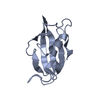


 PDBj
PDBj Assembly


 Mass: 35.453 Da / Num. of mol.: 2 / Source method: obtained synthetically / Formula: Cl
 Mass: 126.904 Da / Num. of mol.: 10 / Source method: obtained synthetically / Formula: I
 Mass: 18.015 Da / Num. of mol.: 84 / Source method: isolated from a natural source / Formula: H2O
 Sample preparation
 / Beamline: 21-ID-G / Wavelength: 0.97856 Å
 Processing