1PX5
 
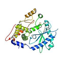
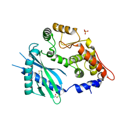 | | Crystal structure of the 2'-specific and double-stranded RNA-activated interferon-induced antiviral protein 2'-5'-oligoadenylate synthetase | | 分子名称: | 2'-5'-oligoadenylate synthetase 1, SULFATE ION | | 著者 | Hartmann, R, Justesen, J, Sarkar, S.N, Sen, G.C, Yee, V.C. | | 登録日 | 2003-07-02 | | 公開日 | 2003-11-25 | | 最終更新日 | 2011-07-13 | | 実験手法 | X-RAY DIFFRACTION (1.74 Å) | | 主引用文献 | Crystal structure of the 2'-specific and double-stranded RNA-activated interferon-induced antiviral protein 2'-5'-oligoadenylate synthetase
Mol.Cell, 12, 2003
|
|
2N4I
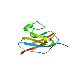 | | The solution structure of Skint-1, a critical determinant of dendritic epidermal gamma-delta T cell selection | | 分子名称: | Selection and upkeep of intraepithelial T-cells protein 1 | | 著者 | Salim, M, Knowles, T.J, Hart, R, Mohammed, F, Woodward, M.J, Willcox, C.R, Overduin, M, Hayday, A.C, Willcox, B.E. | | 登録日 | 2015-06-18 | | 公開日 | 2016-03-02 | | 最終更新日 | 2016-05-11 | | 実験手法 | SOLUTION NMR | | 主引用文献 | Characterization of a Putative Receptor Binding Surface on Skint-1, a Critical Determinant of Dendritic Epidermal T Cell Selection.
J.Biol.Chem., 291, 2016
|
|
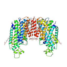
4AC5
 | | Lipidic sponge phase crystal structure of the Bl. viridis reaction centre solved using serial femtosecond crystallography | | 分子名称: | 15-cis-1,2-dihydroneurosporene, BACTERIOCHLOROPHYLL B, BACTERIOPHEOPHYTIN B, ... | | 著者 | Johansson, L.C, Arnlund, D, White, T.A, Katona, G, DePonte, D.P, Weierstall, U, Doak, R.B, Shoeman, R.L, Lomb, L, Malmerberg, E, Davidsson, J, Nass, K, Liang, M, Andreasson, J, Aquila, A, Bajt, S, Barthelmess, M, Barty, A, Bogan, M.J, Bostedt, C, Bozek, J.D, Caleman, C, Coffee, R, Coppola, N, Ekeberg, T, Epp, S.W, Erk, B, Fleckenstein, H, Foucar, L, Graafsma, H, Gumprecht, L, Hajdu, J, Hampton, C.Y, Hartmann, R, Hartmann, A, Hauser, G, Hirsemann, H, Holl, P, Hunter, M.S, Kassemeyer, S, Kimmel, N, Kirian, R.A, Maia, F.R.N.C, Marchesini, S, Martin, A.V, Reich, C, Rolles, D, Rudek, B, Rudenko, A, Schlichting, I, Schulz, J, Seibert, M.M, Sierra, R, Soltau, H, Starodub, D, Stellato, F, Stern, S, Struder, L, Timneanu, N, Ullrich, J, Wahlgren, W.Y, Wang, X, Weidenspointner, G, Wunderer, C, Fromme, P, Chapman, H.N, Spence, J.C.H, Neutze, R. | | 登録日 | 2011-12-14 | | 公開日 | 2012-02-15 | | 最終更新日 | 2023-12-20 | | 実験手法 | X-RAY DIFFRACTION (8.2 Å) | | 主引用文献 | Lipidic Phase Membrane Protein Serial Femtosecond Crystallography.
Nat.Methods, 9, 2012
|
|
7ZGO
 | | Cryo-EM structure of human NKCC1 (TM domain) | | 分子名称: | (2S)-3-(hexadecanoyloxy)-2-[(9Z)-octadec-9-enoyloxy]propyl 2-(trimethylammonio)ethyl phosphate, CHLORIDE ION, CHOLESTEROL HEMISUCCINATE, ... | | 著者 | Nissen, P, Fenton, R, Neumann, C, Lindtoft Rosenbaek, L, Kock Flygaard, R, Habeck, M, Lykkegaard Karlsen, J, Wang, Y, Lindorff-Larsen, K, Gad, H, Hartmann, R, Lyons, J. | | 登録日 | 2022-04-04 | | 公開日 | 2022-10-05 | | 最終更新日 | 2023-04-19 | | 実験手法 | ELECTRON MICROSCOPY (2.55 Å) | | 主引用文献 | Cryo-EM structure of the human NKCC1 transporter reveals mechanisms of ion coupling and specificity.
Embo J., 41, 2022
|
|
3PCQ
 | | Femtosecond X-ray protein Nanocrystallography | | 分子名称: | 1,2-DIPALMITOYL-PHOSPHATIDYL-GLYCEROLE, 1,2-DISTEAROYL-MONOGALACTOSYL-DIGLYCERIDE, BETA-CAROTENE, ... | | 著者 | Chapman, H.N, Fromme, P, Barty, A, White, T.A, Kirian, R.A, Aquila, A, Hunter, M.S, Schulz, J, Deponte, D.P, Weierstall, U, Doak, R.B, Maia, F.R.N.C, Martin, A.V, Schlichting, I, Lomb, L, Coppola, N, Shoeman, R.L, Epp, S.W, Hartmann, R, Rolles, D, Rudenko, A, Foucar, L, Kimmel, N, Weidenspointner, G, Holl, P, Liang, M, Barthelmess, M, Caleman, C, Boutet, S, Bogan, M.J, Krzywinski, J, Bostedt, C, Bajt, S, Gumprecht, L, Rudek, B, Erk, B, Schmidt, C, Homke, A, Reich, C, Pietschner, D, Struder, L, Hauser, G, Gorke, H, Ullrich, J, Herrmann, S, Schaller, G, Schopper, F, Soltau, H, Kuhnel, K.-U, Messerschmidt, M, Bozek, J.D, Hau-Riege, S.P, Frank, M, Hampton, C.Y, Sierra, R, Starodub, D, Williams, G.J, Hajdu, J, Timneanu, N, Seibert, M.M, Andreasson, J, Rocker, A, Jonsson, O, Svenda, M, Stern, S, Nass, K, Andritschke, R, Schroter, C.-D, Krasniqi, F, Bott, M, Schmidt, K.E, Wang, X, Grotjohann, I, Holton, J.M, Barends, T.R.M, Neutze, R, Marchesini, S, Fromme, R, Schorb, S, Rupp, D, Adolph, M, Gorkhover, T, Andersson, I, Hirsemann, H, Potdevin, G, Graafsma, H, Nilsson, B, Spence, J.C.H. | | 登録日 | 2010-10-21 | | 公開日 | 2011-02-02 | | 最終更新日 | 2023-09-06 | | 実験手法 | X-RAY DIFFRACTION (8.984 Å) | | 主引用文献 | Femtosecond X-ray protein nanocrystallography.
Nature, 470, 2011
|
|
2YON
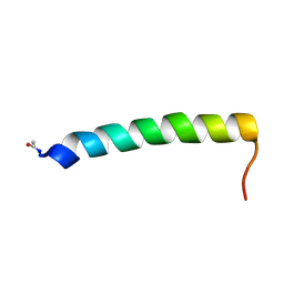 | | Solution NMR structure of the C-terminal extension of two bacterial light, oxygen, voltage (LOV) photoreceptor proteins from Pseudomonas putida | | 分子名称: | SENSORY BOX PROTEIN | | 著者 | Rani, R, Hartmann, R, Lecher, J, Krauss, U, Jaeger, K, Willbold, D. | | 登録日 | 2012-10-25 | | 公開日 | 2013-07-10 | | 最終更新日 | 2019-10-23 | | 実験手法 | SOLUTION NMR | | 主引用文献 | Conservation of Dark Recovery Kinetic Parameters and Structural Features in the Pseudomonadaceae "Short" Light, Oxygen, Voltage (Lov) Protein Family: Implications for the Design of Lov-Based Optogenetic Tools.
Biochemistry, 52, 2013
|
|
2YOM
 | | Solution NMR structure of the C-terminal extension of two bacterial light, oxygen, voltage (LOV) photoreceptor proteins from Pseudomonas putida | | 分子名称: | SENSORY BOX PROTEIN | | 著者 | Rani, R, Lecher, J, Hartmann, R, Krauss, U, Jaeger, K, Willbold, D. | | 登録日 | 2012-10-25 | | 公開日 | 2013-07-10 | | 最終更新日 | 2019-10-23 | | 実験手法 | SOLUTION NMR | | 主引用文献 | Conservation of Dark Recovery Kinetic Parameters and Structural Features in the Pseudomonadaceae "Short" Light, Oxygen, Voltage (Lov) Protein Family: Implications for the Design of Lov-Based Optogenetic Tools.
Biochemistry, 52, 2013
|
|
4XQ7
 | | The crystal structure of the OAS-like domain (OLD) of human OASL | | 分子名称: | 2'-5'-oligoadenylate synthase-like protein | | 著者 | Ibsen, M.S, Gad, H.H, Andersen, L.L, Hornung, V, Julkunen, I, Sarkar, S.N, Hartmann, R. | | 登録日 | 2015-01-19 | | 公開日 | 2015-04-22 | | 最終更新日 | 2024-01-10 | | 実験手法 | X-RAY DIFFRACTION (1.6 Å) | | 主引用文献 | Structural and functional analysis reveals that human OASL binds dsRNA to enhance RIG-I signaling.
Nucleic Acids Res., 43, 2015
|
|
3L9J
 | | Selection of a novel highly specific TNFalpha antagonist: Insight from the crystal structure of the antagonist-TNFalpha complex | | 分子名称: | MAGNESIUM ION, TNFalpha, Tumor necrosis factor, ... | | 著者 | Byla, P, Andersen, M.H, Thogersen, H.C, Gad, H.H, Hartmann, R. | | 登録日 | 2010-01-05 | | 公開日 | 2010-02-23 | | 最終更新日 | 2023-11-01 | | 実験手法 | X-RAY DIFFRACTION (2.1 Å) | | 主引用文献 | Selection of a novel and highly specific TNF{alpha} antagonist: insight from the crystal structure of the antagonist-TNF{alpha} complex
J.Biol.Chem., 285, 2010
|
|
4O6K
 | | The crystal structure of zebrafish IL-22 | | 分子名称: | Interleukin 22 | | 著者 | Siupka, P, Hamming, O.J, Fretaud, M, Luftalla, G, Levraud, J.P, Hartmann, R. | | 登録日 | 2013-12-21 | | 公開日 | 2014-09-10 | | 最終更新日 | 2018-05-23 | | 実験手法 | X-RAY DIFFRACTION (2.1 Å) | | 主引用文献 | The crystal structure of zebrafish IL-22 reveals an evolutionary, conserved structure highly similar to that of human IL-22.
Genes Immun., 15, 2014
|
|
4NZD
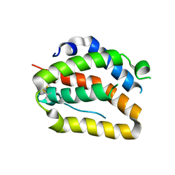
 | | Interleukin 21 receptor | | 分子名称: | 1,2-ETHANEDIOL, CHLORIDE ION, Interleukin-21 receptor, ... | | 著者 | Hamming, O.T, Kang, L, Siupka, P, Gad, H.H, Hartmann, R. | | 登録日 | 2013-12-12 | | 公開日 | 2014-12-17 | | 最終更新日 | 2020-07-29 | | 実験手法 | X-RAY DIFFRACTION (2.75 Å) | | 主引用文献 | Interleukin 21 receptor structure and function
To be Published
|
|
3TGX
 | | IL-21:IL21R complex | | 分子名称: | Interleukin-21, Interleukin-21 receptor, NICKEL (II) ION, ... | | 著者 | Hamming, O.J, Kang, L, Svenson, A, Karlsen, J.L, Rahbek-Nielsen, H, Paludan, S.R, Hjort, S.A, Bondensgaard, K, Hartmann, R. | | 登録日 | 2011-08-18 | | 公開日 | 2012-02-15 | | 最終更新日 | 2023-12-27 | | 実験手法 | X-RAY DIFFRACTION (2.8 Å) | | 主引用文献 | The crystal structure of the interleukin 21 receptor bound to interleukin 21 reveals that a sugar chain interacting with the WSXWS motif is an integral part of the interleukin 21 receptor.
J.Biol.Chem., 2012
|
|
3PIW
 | |
3PIV
 | | Zebrafish interferon 1 | | 分子名称: | Interferon, NICKEL (II) ION | | 著者 | Hamming, O.J, Hartmann, R, Lutfalla, G, Levraud, J.-P. | | 登録日 | 2010-11-08 | | 公開日 | 2011-07-20 | | 最終更新日 | 2018-03-07 | | 実験手法 | X-RAY DIFFRACTION (2.086 Å) | | 主引用文献 | Crystal Structure of Zebrafish Interferons I and II Reveals Conservation of Type I Interferon Structure in Vertebrates.
J.Virol., 85, 2011
|
|
3HEQ
 | | Human prion protein variant D178N with M129 | | 分子名称: | CADMIUM ION, Major prion protein | | 著者 | Lee, S, Antony, L, Hartmann, R, Knaus, K.J, Surewicz, K, Surewicz, W.K, Yee, V.C. | | 登録日 | 2009-05-10 | | 公開日 | 2010-01-12 | | 最終更新日 | 2021-10-13 | | 実験手法 | X-RAY DIFFRACTION (1.8 Å) | | 主引用文献 | Conformational diversity in prion protein variants influences intermolecular beta-sheet formation.
Embo J., 29, 2010
|
|
3HES
 | | Human prion protein variant F198S with M129 | | 分子名称: | CADMIUM ION, Major prion protein | | 著者 | Lee, S, Antony, L, Hartmann, R, Knaus, K.J, Surewicz, K, Surewicz, W.K, Yee, V.C. | | 登録日 | 2009-05-10 | | 公開日 | 2010-01-12 | | 最終更新日 | 2021-10-13 | | 実験手法 | X-RAY DIFFRACTION (2 Å) | | 主引用文献 | Conformational diversity in prion protein variants influences intermolecular beta-sheet formation.
Embo J., 29, 2010
|
|
3HHC
 | |
3HAF
 | | Human prion protein variant V129 domain swapped dimer | | 分子名称: | CADMIUM ION, CHLORIDE ION, Major prion protein | | 著者 | Lee, S, Antony, L, Hartmann, R, Knaus, K.J, Surewicz, K, Surewicz, W.K, Yee, V.C. | | 登録日 | 2009-05-01 | | 公開日 | 2010-01-12 | | 最終更新日 | 2017-11-01 | | 実験手法 | X-RAY DIFFRACTION (2.26 Å) | | 主引用文献 | Conformational diversity in prion protein variants influences intermolecular beta-sheet formation.
Embo J., 29, 2010
|
|
3HAK
 | | Human prion protein variant V129 | | 分子名称: | Major prion protein | | 著者 | Lee, S, Antony, L, Hartmann, R, Knaus, K.J, Surewicz, K, Surewicz, W.K, Yee, V.C. | | 登録日 | 2009-05-01 | | 公開日 | 2010-01-12 | | 最終更新日 | 2017-11-01 | | 実験手法 | X-RAY DIFFRACTION (1.8 Å) | | 主引用文献 | Conformational diversity in prion protein variants influences intermolecular beta-sheet formation.
Embo J., 29, 2010
|
|
3HJX
 | | Human prion protein variant D178N with V129 | | 分子名称: | CADMIUM ION, CHLORIDE ION, Major prion protein | | 著者 | Lee, S, Antony, L, Hartmann, R, Knaus, K.J, Surewicz, K, Surewicz, W.K, Yee, V.C. | | 登録日 | 2009-05-22 | | 公開日 | 2010-01-12 | | 最終更新日 | 2021-10-13 | | 実験手法 | X-RAY DIFFRACTION (2 Å) | | 主引用文献 | Conformational diversity in prion protein variants influences intermolecular beta-sheet formation.
Embo J., 29, 2010
|
|
3HER
 | | Human prion protein variant F198S with V129 | | 分子名称: | CADMIUM ION, Major prion protein | | 著者 | Lee, S, Antony, L, Hartmann, R, Knaus, K.J, Surewicz, K, Surewicz, W.K, Yee, V.C. | | 登録日 | 2009-05-10 | | 公開日 | 2010-01-12 | | 最終更新日 | 2021-10-13 | | 実験手法 | X-RAY DIFFRACTION (1.85 Å) | | 主引用文献 | Conformational diversity in prion protein variants influences intermolecular beta-sheet formation.
Embo J., 29, 2010
|
|
3HJ5
 | | Human prion protein variant V129 domain swapped dimer | | 分子名称: | Major prion protein | | 著者 | Lee, S, Antony, L, Hartmann, R, Knaus, K.J, Surewicz, K, Surewicz, W.K, Yee, V.C. | | 登録日 | 2009-05-20 | | 公開日 | 2010-01-12 | | 最終更新日 | 2017-11-01 | | 実験手法 | X-RAY DIFFRACTION (3.1 Å) | | 主引用文献 | Conformational diversity in prion protein variants influences intermolecular beta-sheet formation.
Embo J., 29, 2010
|
|
4BII
 | |
2QMV
 | |
5IFH
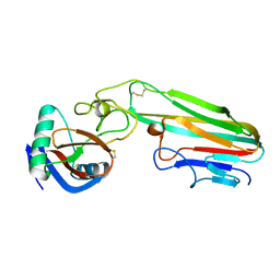
 | | Crystal structure of the BCR Fab fragment from subset #2 case P11475 | | 分子名称: | Heavy chain of the Fab fragment from BCR derived from the P11475 CLL clone, Light chain of the Fab fragment from BCR derived from the P11475 CLL clone | | 著者 | Minici, C, Degano, M. | | 登録日 | 2016-02-26 | | 公開日 | 2017-03-08 | | 最終更新日 | 2024-01-10 | | 実験手法 | X-RAY DIFFRACTION (2.293 Å) | | 主引用文献 | Distinct homotypic B-cell receptor interactions shape the outcome of chronic lymphocytic leukaemia.
Nat Commun, 8, 2017
|
|


