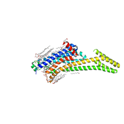
10AD
 
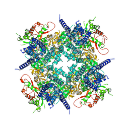 | | Cryo-EM structure of the human BK channel bound to the agonist NS1619 | | Descriptor: | 3-[2-oxidanyl-5-(trifluoromethyl)phenyl]-6-(trifluoromethyl)-1~{H}-benzimidazol-2-one, CALCIUM ION, Isoform 5 of Calcium-activated potassium channel subunit alpha-1, ... | | Authors: | Gonzalez-Sanabria, N, Contreras, G.F, Perozo, E, Latorre, R. | | Deposit date: | 2026-01-08 | | Release date: | 2026-02-04 | | Last modified: | 2026-02-11 | | Method: | ELECTRON MICROSCOPY (3.44 Å) | | Cite: | The BK channel-NS1619 agonist complex reveals molecular insights into allosteric activation gating.
Proc.Natl.Acad.Sci.USA, 123, 2026
|
|
10AH
 | |
10AI
 | |
10AJ
 | | Crystal Structure of Human WRN helicase with compound 1 | | Descriptor: | 1,2-ETHANEDIOL, 4-(4-methyl-4H-1,2,4-triazol-3-yl)-1-[4-(1H-pyrrol-1-yl)benzene-1-sulfonyl]piperidine, Bifunctional 3'-5' exonuclease/ATP-dependent helicase WRN, ... | | Authors: | Toms, A.V, Caravella, J.A, Sitnikov, N, Bartels, F, Svensson, R, Jacques O'Hagan, S, Borthwick, J, Campos, S, Yin, Y, Zhao, X, Li, L, Talbot, E, Kong, H, Freund, R.R.A, Browning, B, Genung, N.E, Carreiro, S, Brennan, D, Graves, A.P, Loh, C, Tummino, P, Edmonson, S.E, Li, D. | | Deposit date: | 2026-01-08 | | Release date: | 2026-05-13 | | Last modified: | 2026-06-10 | | Method: | X-RAY DIFFRACTION (2.42 Å) | | Cite: | Design of Cyclic Vinyl Sulfones as WRN Covalent Inhibitors from Noncovalent Binders.
J.Med.Chem., 69, 2026
|
|
10AK
 | | Crystal Structure of Human WRN helicase with compound 4 | | Descriptor: | 1,2-ETHANEDIOL, Bifunctional 3'-5' exonuclease/ATP-dependent helicase WRN, CHLORIDE ION, ... | | Authors: | Toms, A.V, Caravella, J.A, Sitnikov, N, Bartels, F, Svensson, R, Jacques O'Hagan, S, Borthwick, J, Yin, Y, Zhoa, X, Li, L, Liu, R, Talbot, E, Kong, H, Freund, R.R.A, Browning, B, Genung, N, Carreiro, S, Brennan, D, Graves, A.P, Loh, C, Tummino, P, Edmondson, S.D, Li, D. | | Deposit date: | 2026-01-08 | | Release date: | 2026-05-13 | | Last modified: | 2026-06-10 | | Method: | X-RAY DIFFRACTION (1.37 Å) | | Cite: | Design of Cyclic Vinyl Sulfones as WRN Covalent Inhibitors from Noncovalent Binders.
J.Med.Chem., 69, 2026
|
|
10AP
 | | Crystal Structure of Human WRN helicase with compound 26 | | Descriptor: | (2R)-N-[(3R)-1,1-dioxo-1lambda~6~-thiolan-3-yl]-N-{[2-(2-hydroxypropan-2-yl)pyridin-4-yl]methyl}-2-methoxy-2-[(1M)-3,3',4'-trifluoro[1,1'-biphenyl]-4-yl]acetamide, 1,2-ETHANEDIOL, ADENOSINE-5'-TRIPHOSPHATE, ... | | Authors: | Toms, A.V, Caravella, J.A, Sitnikov, N, Bartels, F, Svensson, R, Jacques O'Hagan, S, Borthwick, J, Campos, S, Yin, Y, Zhao, X, Li, L, Talbot, E, Kong, H, Freund, R.R.A, Browning, B, Genung, N.E, Carreiro, S, Brennan, D, Graves, A.P, Loh, C, Tummino, P, Edmonson, S.D, Li, D. | | Deposit date: | 2026-01-08 | | Release date: | 2026-05-13 | | Last modified: | 2026-06-10 | | Method: | X-RAY DIFFRACTION (2.58 Å) | | Cite: | Design of Cyclic Vinyl Sulfones as WRN Covalent Inhibitors from Noncovalent Binders.
J.Med.Chem., 69, 2026
|
|
10AY
 | | Cryo-EM structure of CRBN-DDB1 in complex with HBS1L and TNG961 | | Descriptor: | DNA damage-binding protein 1, HBS1-like protein, N-{6-[(3R)-2,6-dioxopiperidin-3-yl]naphthalen-1-yl}-N'-{2-[6-(trifluoromethyl)-1-benzothiophen-2-yl]propan-2-yl}urea, ... | | Authors: | Whittington, D.A. | | Deposit date: | 2026-01-09 | | Release date: | 2026-04-22 | | Last modified: | 2026-05-06 | | Method: | ELECTRON MICROSCOPY (2.9 Å) | | Cite: | TNG961 is a selective oral HBS1L molecular glue degrader for the treatment of FOCAD-deleted cancers.
Cancer Discov, 2026
|
|
10BT
 | | X-ray Crystal Structure of A High-Affinity Monoclonal Antibody Sequesters Xylazine | | Descriptor: | Antibody Heavy Chain, Antibody Light Chain, CHLORIDE ION, ... | | Authors: | Shi, K, Moller, N, Aihara, H. | | Deposit date: | 2026-01-10 | | Release date: | 2026-06-10 | | Method: | X-RAY DIFFRACTION (1.99 Å) | | Cite: | Discovery, Structural Characterization, and Preclinical Evaluation of Monoclonal Antibodies against Xylazine Poisoning
Acs Pharmacol Transl Sci, 2026
|
|
10FZ
 | | 30S ribosomal subunit from E. coli missing the gene encoding for the 16S rRNA 2'-O-methyltransferase RsmI | | Descriptor: | 16S rRNA, MAGNESIUM ION, Small ribosomal subunit protein bS16, ... | | Authors: | Barmada, M.I, Nandi, S, Conn, G.L. | | Deposit date: | 2026-01-18 | | Release date: | 2026-03-18 | | Last modified: | 2026-04-08 | | Method: | ELECTRON MICROSCOPY (2.91 Å) | | Cite: | Mechanism of 30S subunit recognition and modification by the conserved bacterial ribosomal RNA methyltransferase RsmI.
Proc.Natl.Acad.Sci.USA, 123, 2026
|
|
10HM
 | |
10HY
 | | Structure of CHK1 10-pt. mutant complex with macrocyclic LRRK2 inhibitor compound 1 ((11R)-8-chloro-3,11-dimethyl-2-(oxan-4-yl)-2,4,10,11,12,13-hexahydro-9,5-(azeno)pyrazolo[3,4-b][1,4,6,10]oxatriazacyclotridecine) | | Descriptor: | (11R)-8-chloro-3,11-dimethyl-2-(oxan-4-yl)-2,4,10,11,12,13-hexahydro-9,5-(azeno)pyrazolo[3,4-b][1,4,6,10]oxatriazacyclotridecine, Serine/threonine-protein kinase Chk1 | | Authors: | Palte, R.L, Yu, E.C, Zhou, H. | | Deposit date: | 2026-01-21 | | Release date: | 2026-04-08 | | Last modified: | 2026-04-22 | | Method: | X-RAY DIFFRACTION (2.03 Å) | | Cite: | Discovery of Potent, Selective, CNS-Penetrant Macrocyclic LRRK2 Inhibitors for the Treatment of Parkinson's Disease.
J.Med.Chem., 69, 2026
|
|
10HZ
 | | Structure of CHK1 10-pt. mutant complex with macrocyclic LRRK2 inhibitor compound 7 ((10aS,13aS)-3-cyclobutyl-1-methyl-8-(trifluoromethyl)-3,4,10a,11,13a,14-hexahydro-10H,13H-9,5-(azeno)furo[3,4-k]pyrazolo[4,3-b][1,4,6,10]oxatriazacyclotridecine) | | Descriptor: | (10aS,13aS)-3-cyclobutyl-1-methyl-8-(trifluoromethyl)-3,4,10a,11,13a,14-hexahydro-10H,13H-9,5-(azeno)furo[3,4-k]pyrazolo[4,3-b][1,4,6,10]oxatriazacyclotridecine, Serine/threonine-protein kinase Chk1 | | Authors: | Palte, R.L, Yu, E.C, Zhou, H. | | Deposit date: | 2026-01-21 | | Release date: | 2026-04-08 | | Last modified: | 2026-04-22 | | Method: | X-RAY DIFFRACTION (1.666 Å) | | Cite: | Discovery of Potent, Selective, CNS-Penetrant Macrocyclic LRRK2 Inhibitors for the Treatment of Parkinson's Disease.
J.Med.Chem., 69, 2026
|
|
10IA
 | | Structure of CHK1 10-pt. mutant complex with macrocyclic LRRK2 inhibitor compound 12 ((10aS,13aS)-3-cyclopropyl-1-methyl-8-(trifluoromethyl)-3,4,10a,11,13a,14-hexahydro-10H,13H-9,5-(azeno)furo[3,4-k]pyrazolo[4,3-b][1,4,6,10]oxatriazacyclotridecine) | | Descriptor: | (10aS,13aS)-3-cyclopropyl-1-methyl-8-(trifluoromethyl)-3,4,10a,11,13a,14-hexahydro-10H,13H-9,5-(azeno)furo[3,4-k]pyrazolo[4,3-b][1,4,6,10]oxatriazacyclotridecine, Serine/threonine-protein kinase Chk1 | | Authors: | Palte, R.L, Yu, E.C, Zhou, H. | | Deposit date: | 2026-01-21 | | Release date: | 2026-04-08 | | Last modified: | 2026-04-22 | | Method: | X-RAY DIFFRACTION (1.739 Å) | | Cite: | Discovery of Potent, Selective, CNS-Penetrant Macrocyclic LRRK2 Inhibitors for the Treatment of Parkinson's Disease.
J.Med.Chem., 69, 2026
|
|
10IC
 | | Rhesus rotavirus (consensus structure at 4.7 Angstrom resolution from cryo-ET) | | Descriptor: | 2-acetamido-2-deoxy-beta-D-glucopyranose, CALCIUM ION, CHLORIDE ION, ... | | Authors: | de Sautu, M, Leistner, C, Kirchhausen, T, Jenni, S, Harrison, S.C. | | Deposit date: | 2026-01-21 | | Release date: | 2026-03-04 | | Method: | ELECTRON MICROSCOPY (4.7 Å) | | Cite: | Mechanism of membrane perforation in rotavirus cell entry.
Biorxiv, 2026
|
|
10JT
 | | CRYSTAL STRUCTURE OF KIRSTEN RAT SARCOMA G12C COMPLEXED WITH GMPPNP AND COVALENTLY BOUND TO 1-[(2R,3R)-3-{[(7P)-7-(8-ethynyl-7-fluoronaphthalen-1-yl)-8-fluoro-2-{ [(2R,4R,7aS)-2-fluorotetrahydro-1H-pyrrolizin-7a(5H)-yl]methoxy}pyrido[4,3-d] pyrimidin-4-yl](methyl)amino}-2-methylpyrrolidin-1-yl]-3-(pyrazin-2-yl)propan-1-one | | Descriptor: | 1-[(2R,3R)-3-{[(7P)-7-(8-ethynyl-7-fluoronaphthalen-1-yl)-8-fluoro-2-{[(2R,4R,7aS)-2-fluorotetrahydro-1H-pyrrolizin-7a(5H)-yl]methoxy}pyrido[4,3-d]pyrimidin-4-yl](methyl)amino}-2-methylpyrrolidin-1-yl]-3-(pyrazin-2-yl)propan-1-one, CALCIUM ION, GUANOSINE-5'-DIPHOSPHATE, ... | | Authors: | Sheriff, S. | | Deposit date: | 2026-01-22 | | Release date: | 2026-03-04 | | Last modified: | 2026-03-18 | | Method: | X-RAY DIFFRACTION (1.489 Å) | | Cite: | Optimization of Covalent Warhead Trajectory for KRAS G12C Active-State Inhibition.
J.Med.Chem., 69, 2026
|
|
10KT
 | | Crystal structure of A2A adenosine receptor A2AR-bRIL in complex with Compound50 | | Descriptor: | (2R)-2,3-dihydroxypropyl (9Z)-octadec-9-enoate, (2S)-2,3-dihydroxypropyl (9Z)-octadec-9-enoate, (4%{S})-2-[(3%{R},6%{S})-1-(1-ethyl-1%{H}-pyrazol-4-yl)-6-methylpiperidin-3-yl]-7-methoxy[1,2,4]triazolo[1,5-%{c}]quinazolin-5-amine, ... | | Authors: | Krishnamurthy, H. | | Deposit date: | 2026-01-25 | | Release date: | 2026-05-20 | | Last modified: | 2026-05-27 | | Method: | X-RAY DIFFRACTION (2.59 Å) | | Cite: | Discovery of MK-1088 as a Potent A 2A /A 2B Adenosine Receptor Dual-Antagonist for Cancer Immunotherapy.
J.Med.Chem., 69, 2026
|
|
10KZ
 | | N-Alkyl & N-Aryl Aminopyrazole Spirocarbamates: A Two-Pronged Lead Optimization Strategy to Identify Orally Bioavailable Plasma Kallikrein Inhibitors | | Descriptor: | (3'R)-1'-(1-benzyl-1H-pyrazole-4-carbonyl)-6-chloro-5-fluorospiro[[3,1]benzoxazine-4,3'-piperidin]-2(1H)-one, Plasma kallikrein | | Authors: | Merchant, R.R, Chernyak, N, Lopez, J.A, Sharp, P.P, Mandal, M, He, J, Hruza, A, Rearden, P, Tatosian, D.A, Lin, K, Esmay, J, Yang, S, Cheng, A, Ellsworth, K, Piou, T, Fier, P, Hicks, J, Sinz, C, Ogawa, A. | | Deposit date: | 2026-01-26 | | Release date: | 2026-03-11 | | Last modified: | 2026-04-01 | | Method: | X-RAY DIFFRACTION (1.78 Å) | | Cite: | N ‐Alkyl and N ‐Aryl Aminopyrazole Spirocarbamates: A Two-Pronged Lead Optimization Strategy to Identify Orally Bioavailable Plasma Kallikrein Inhibitors.
Acs Med.Chem.Lett., 17, 2026
|
|
10LR
 | | N-Alkyl & N-Aryl Aminopyrazole Spirocarbamates: A Two-Pronged Lead Optimization Strategy to Identify Orally Bioavailable PlasmaKallikrein Inhibitors complex with Compound 4 ((3'R)-1'-(5-amino-1-benzyl-1H-pyrazole-4-carbonyl)-6-chloro-5-fluorospiro[[3,1]benzoxazine-4,3'-piperidin]-2(1H)-one) | | Descriptor: | (3'R)-1'-(5-amino-1-benzyl-1H-pyrazole-4-carbonyl)-6-chloro-5-fluorospiro[[3,1]benzoxazine-4,3'-piperidin]-2(1H)-one, 2-(N-MORPHOLINO)-ETHANESULFONIC ACID, 2-acetamido-2-deoxy-beta-D-glucopyranose, ... | | Authors: | Merchant, R.R, Chernyak, N, Lopez, J.A, Sharp, P.P, Mandal, M, He, J, Hruza, A, Rearden, P, Tatosian, D.A, Lin, K, Esmay, J, Yang, S, Cheng, A, Ellsworth, K, Piou, T, Fier, P, Hicks, J, Sinz, C, Ogawa, A. | | Deposit date: | 2026-01-27 | | Release date: | 2026-03-11 | | Last modified: | 2026-04-01 | | Method: | X-RAY DIFFRACTION (1.583 Å) | | Cite: | N ‐Alkyl and N ‐Aryl Aminopyrazole Spirocarbamates: A Two-Pronged Lead Optimization Strategy to Identify Orally Bioavailable Plasma Kallikrein Inhibitors.
Acs Med.Chem.Lett., 17, 2026
|
|
10LX
 | | High Stable Quinonoid Intermediate of Human Ornithine Aminotransferase Complexed with (1R,4S)-4-Amino-3-(trifluoromethyl)cyclopent-2-ene-1-carboxylic Acid | | Descriptor: | (1S,4R)-4-(difluoromethyl)-3-[({3-hydroxy-2-methyl-5-[(phosphonooxy)methyl]pyridin-4-yl}methyl)amino]cyclopent-2-ene-1-carboxylic acid, Ornithine aminotransferase, mitochondrial | | Authors: | Vargas, A.L, Liu, D. | | Deposit date: | 2026-01-27 | | Release date: | 2026-05-20 | | Method: | X-RAY DIFFRACTION (1.83 Å) | | Cite: | Inactivation of ornithine aminotransferase by (1 R ,4 S )-4-Amino-3-(trifluoromethyl)cyclopent-2-ene-1-carboxylic acid via a stable quinonoid intermediate.
Med.Chem.Res., 35, 2026
|
|
10ML
 | |
10MN
 | | Crystal structure of human OGG1 (WT) in a product bound state in the presence of the agonist F51 | | Descriptor: | (2Z)-4-hydroxypenta-2,4-dienal, 1-[(2S)-2-(benzyloxy)-2-(2,4-dichlorophenyl)ethyl]-1H-imidazole, DNA (5'-D(*TP*AP*GP*AP*GP*TP*C*(PUA))-3'), ... | | Authors: | Syed, A, Arvai, A.S, Lloyd, R.S, Tainer, J.A. | | Deposit date: | 2026-01-27 | | Release date: | 2026-07-15 | | Method: | X-RAY DIFFRACTION (2.1 Å) | | Cite: | Crystal structure of human OGG1 (WT) in a product bound state in the presence of the agonist F51
To Be Published
|
|
10MT
 | | C2 symmetry expanded and subtracted 20S Proteasome, Blm10, Fub1 Complex Halfmer | | Descriptor: | Proteasome activator BLM10, Proteasome subunit alpha type-1, Proteasome subunit alpha type-2, ... | | Authors: | Walsh Jr, R.M, Rawson, S, Fermin Perez, E, Venclovaite, U, Hanna, J. | | Deposit date: | 2026-01-28 | | Release date: | 2026-07-22 | | Method: | ELECTRON MICROSCOPY (3.7 Å) | | Cite: | Blm10 and PI31 Compromise a Failsafe Mechanism for Proteasome Inhibition
To Be Published
|
|
10MV
 | | N-Alkyl & N-Aryl Aminopyrazole Spirocarbamates: A Two-Pronged Lead Optimization Strategy to Identify Orally Bioavailable Plasma Kallikrein Inhibitors complex with Compound 15 ((3'R)-1'-(5-amino-1-phenyl-1H-pyrazole-4-carbonyl)-6-chloro-5-fluorospiro[[3,1]benzoxazine-4,3'-piperidin]-2(1H)-one) | | Descriptor: | (3'R)-1'-(5-amino-1-phenyl-1H-pyrazole-4-carbonyl)-6-chloro-5-fluorospiro[[3,1]benzoxazine-4,3'-piperidin]-2(1H)-one, 2-(N-MORPHOLINO)-ETHANESULFONIC ACID, 2-acetamido-2-deoxy-beta-D-glucopyranose, ... | | Authors: | Merchant, R.R, Chernyak, N, Lopez, J.A, Sharp, P.P, Mandal, M, He, J, Hruza, A, Rearden, P, Tatosian, D.A, Lin, K, Esmay, J, Yang, S, Cheng, A, Ellsworth, K, Ogawa, A, Piou, T, Fier, P, Hicks, J, Sinz, C, Ogawa, A. | | Deposit date: | 2026-01-28 | | Release date: | 2026-04-01 | | Method: | X-RAY DIFFRACTION (1.66 Å) | | Cite: | N ‐Alkyl and N ‐Aryl Aminopyrazole Spirocarbamates: A Two-Pronged Lead Optimization Strategy to Identify Orally Bioavailable Plasma Kallikrein Inhibitors.
Acs Med.Chem.Lett., 17, 2026
|
|
10MW
 | | N-Alkyl & N-Aryl Aminopyrazole Spirocarbamates: A Two-Pronged Lead Optimization Strategy to Identify Orally Bioavailable Plasma Kallikrein Inhibitors compound 25 ((3'R)-1'-{(1P)-5-amino-1-[2-(trifluoromethoxy)phenyl]-1H-pyrazole-4-carbonyl}-6-chloro-5-fluorospiro[[3,1]benzoxazine-4,3'-piperidin]-2(1H)-one) | | Descriptor: | (3'R)-1'-{(1P)-5-amino-1-[2-(trifluoromethoxy)phenyl]-1H-pyrazole-4-carbonyl}-6-chloro-5-fluorospiro[[3,1]benzoxazine-4,3'-piperidin]-2(1H)-one, 2-(N-MORPHOLINO)-ETHANESULFONIC ACID, 2-acetamido-2-deoxy-beta-D-glucopyranose, ... | | Authors: | Merchant, R.R, Chernyak, N, Lopez, J.A, Sharp, P.P, Mandal, M, Je, J, Hruza, A, Rearden, P, Tatosian, D.A, Lin, K, Esmay, J, Yang, S, Cheng, A, Ellsworth, K, Poiou, T, Fier, P, Hicks, J, Sinz, C, Ogawa, A. | | Deposit date: | 2026-01-28 | | Release date: | 2026-03-11 | | Last modified: | 2026-04-01 | | Method: | X-RAY DIFFRACTION (1.62 Å) | | Cite: | N ‐Alkyl and N ‐Aryl Aminopyrazole Spirocarbamates: A Two-Pronged Lead Optimization Strategy to Identify Orally Bioavailable Plasma Kallikrein Inhibitors.
Acs Med.Chem.Lett., 17, 2026
|
|
10NU
 | | Structure of kRas G12C bound to Inhibitor 13ab | | Descriptor: | 1-((2R,5S)-4-((S)-6-chloro-7-(1,6-dimethyl-1H-indazol-7-yl)-8-fluoro-2-(((S)-1-methylpyrrolidin-2-yl)methoxy)quinazolin-4-yl)-2,5-dimethylpiperazin-1-yl)prop-2-en-1-one, CALCIUM ION, GLYCEROL, ... | | Authors: | Shaffer, P.L, Milligan, C, Peters, U. | | Deposit date: | 2026-01-29 | | Release date: | 2026-04-15 | | Last modified: | 2026-04-29 | | Method: | X-RAY DIFFRACTION (1.5 Å) | | Cite: | Optimization of Covalent 6-Cyanoquinazoline KRAS G12C Inhibitors for the Treatment of Solid Tumors.
J.Med.Chem., 69, 2026
|
|


