 Movie
Movie Controller


+ Open data

- Basic information
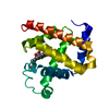
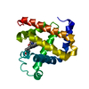
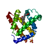
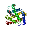
| Entry | Database: PDB / ID: 1h1x | ||||||
|---|---|---|---|---|---|---|---|
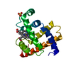
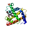
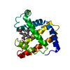
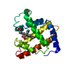
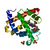
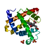
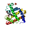
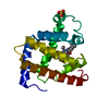
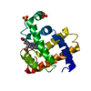
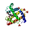
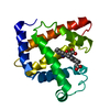
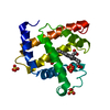
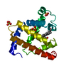
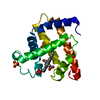
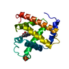
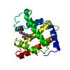
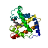
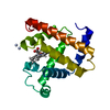
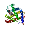
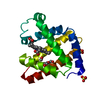
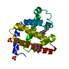
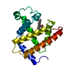
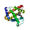
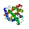
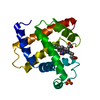
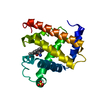
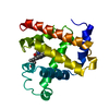
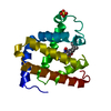
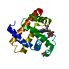
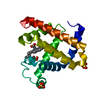
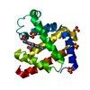
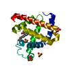
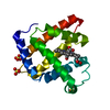
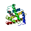
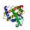
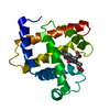
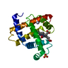
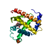
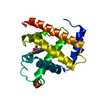
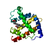
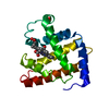
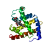
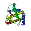
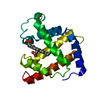
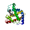
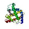
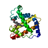
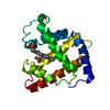
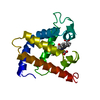
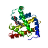
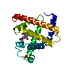
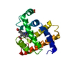
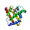
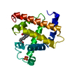
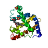
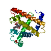
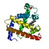
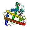
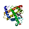
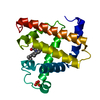
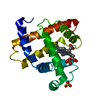
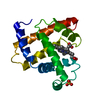
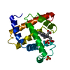
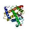
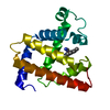
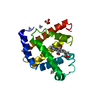
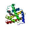
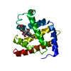
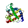
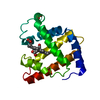
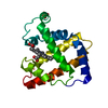
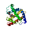
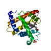
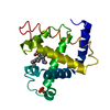
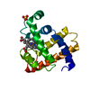
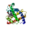
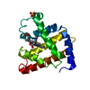
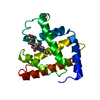
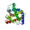
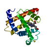
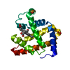
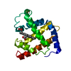
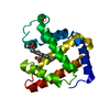
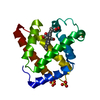
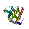
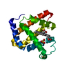
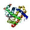
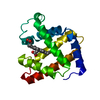
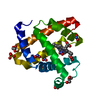
| Title | Sperm whale Myoglobin mutant T67R S92D | ||||||
 Components Components | MYOGLOBIN | ||||||
 Keywords Keywords | OXYGEN TRANSPORT / GLOBIN / PEROXIDASE / OXYGEN STORAGE / HEME / MUSCLE | ||||||
| Function / homology |  Function and homology information Function and homology informationOxidoreductases; Acting on other nitrogenous compounds as donors / nitrite reductase activity / sarcoplasm / Oxidoreductases; Acting on a peroxide as acceptor; Peroxidases / removal of superoxide radicals / oxygen carrier activity / peroxidase activity / oxygen binding / heme binding / extracellular exosome / metal ion binding Similarity search - Function | ||||||
| Biological species |  | ||||||
| Method |  X-RAY DIFFRACTION / SYNCHROTRON / MOLECULAR REPLACEMENT / Resolution: 1.4 Å X-RAY DIFFRACTION / SYNCHROTRON / MOLECULAR REPLACEMENT / Resolution: 1.4 Å | ||||||
 Authors Authors | Zuccotti, S. / Bolognesi, M. | ||||||
 Citation Citation | Journal: Biochem.J. / Year: 2004 Title: Engineering Peroxidase Activity in Myoglobin: The Haem Cavity Structure and Peroxide Activation in the T67R/S92D Mutant and its Derivative Reconstituted with Protohaemin-L-Histidine. Authors: Roncone, R. / Monzani, E. / Murtas, M. / Battaini, G. / Pennati, A. / Sanangelantoni, A.M. / Zuccotti, S. / Bolognesi, M. / Casella, L. #1: Journal: J.Mol.Biol. / Year: 1993Title: High-Resolution Crystal Structures of Distal Histidine Mutants of Sperm Whale Myoglobin Authors: Quillin, M.L. / Arduini, R.M. / Olson, J.S. / Phillips Jr, G.N. | ||||||
| History |
|
- Structure visualization
| Structure viewer | Molecule: MolmilJmol/JSmol |
|---|
- Downloads & links
-Download
| PDBx/mmCIF format | 1h1x.cif.gz | 58.6 KB | Display | PDBx/mmCIF format |
|---|---|---|---|---|
| PDB format | pdb1h1x.ent.gz | 41.6 KB | Display | PDB format |
| PDBx/mmJSON format | 1h1x.json.gz | Tree view | PDBx/mmJSON format | |
| Others |  Other downloads Other downloads |
-Validation report
| Arichive directory | https://data.pdbj.org/pub/pdb/validation_reports/h1/1h1xftp://data.pdbj.org/pub/pdb/validation_reports/h1/1h1x | HTTPS FTP |
|---|
-Related structure data
| Related structure data | 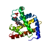 1fcsS S: Starting model for refinement |
|---|---|
| Similar structure data |


-Links
 PDBj
PDBj


- Assembly
| Deposited unit | 
| ||||||||
|---|---|---|---|---|---|---|---|---|---|
| 1 |
| ||||||||
| Unit cell |
|
-Components
| #1: Protein | Mass: 17449.262 Da / Num. of mol.: 1 / Mutation: YES Source method: isolated from a genetically manipulated source Details: MUTATION THR 67 ARG AND SER 92 ASP / Source: (gene. exp.)  |
|---|---|
| #2: Chemical | ChemComp-HEM /   Mass: 616.487 Da / Num. of mol.: 1 / Source method: obtained synthetically / Formula: C34H32FeN4O4 Mass: 616.487 Da / Num. of mol.: 1 / Source method: obtained synthetically / Formula: C34H32FeN4O4 |
| #3: Chemical | ChemComp-CYN /   Mass: 26.017 Da / Num. of mol.: 1 / Source method: obtained synthetically / Formula: CN Mass: 26.017 Da / Num. of mol.: 1 / Source method: obtained synthetically / Formula: CN |
| #4: Chemical | ChemComp-SO4 /   Mass: 96.063 Da / Num. of mol.: 1 / Source method: obtained synthetically / Formula: SO4 Mass: 96.063 Da / Num. of mol.: 1 / Source method: obtained synthetically / Formula: SO4 |
| #5: Water | ChemComp-HOH /  Mass: 18.015 Da / Num. of mol.: 302 / Source method: isolated from a natural source / Formula: H2O Mass: 18.015 Da / Num. of mol.: 302 / Source method: isolated from a natural source / Formula: H2O |
| Compound details | MEMBER OF THE GLOBIN FAMILY SERVING AS RESERVIOR OF OXYGEN IN MUSCLE CELLS ENGINEERED MUTATION THR ...MEMBER OF THE GLOBIN FAMILY SERVING AS RESERVIOR OF OXYGEN IN MUSCLE CELLS ENGINEERED |
| Sequence details | REFERENCE BIOCHIM. BIOPHYS. ACTA 336:318-323(1974) SHOWS CONFLICT AT POSITION 122 OF THE PROTEIN ...REFERENCE BIOCHIM. BIOPHYS. ACTA 336:318-323(1974) SHOWS CONFLICT AT POSITION 122 OF THE PROTEIN SEQUENCE AS INDICATED IN THE DBREF RECORDS BELOW. |
-Experimental details
-Experiment
| Experiment | Method: X-RAY DIFFRACTION / Number of used crystals: 1 |
|---|
- Sample preparation
| Crystal | Density Matthews: 2.34 Å3/Da / Density % sol: 47 % | |||||||||||||||||||||||||||||||||||||||||||||||||
|---|---|---|---|---|---|---|---|---|---|---|---|---|---|---|---|---|---|---|---|---|---|---|---|---|---|---|---|---|---|---|---|---|---|---|---|---|---|---|---|---|---|---|---|---|---|---|---|---|---|---|
| Crystal grow | pH: 9 Details: 3M (NH4)2SO4, 5MM K3FE(CN)6, 20MM TRIS/HCL, PH9.0, DROPLET 5:3, RESERVOIR:PROTEIN (43MG/ML), pH 9.00 | |||||||||||||||||||||||||||||||||||||||||||||||||
| Crystal grow | *PLUS pH: 6 / Method: vapor diffusion, hanging drop | |||||||||||||||||||||||||||||||||||||||||||||||||
| Components of the solutions | *PLUS
|
-Data collection
| Diffraction | Mean temperature: 100 K |
|---|---|
| Diffraction source | Source: SYNCHROTRON / Site: ESRF  / Beamline: ID14-2 / Wavelength: 0.933 / Beamline: ID14-2 / Wavelength: 0.933 |
| Detector | Detector: CCD / Date: Apr 15, 2002 |
| Radiation | Protocol: SINGLE WAVELENGTH / Monochromatic (M) / Laue (L): M / Scattering type: x-ray |
| Radiation wavelength | Wavelength: 0.933 Å / Relative weight: 1 |
| Reflection | Resolution: 1.4→19 Å / Num. obs: 41560 / % possible obs: 99.1 % / Observed criterion σ(I): 3 / Redundancy: 13.51 % / Rmerge(I) obs: 0.057 / Net I/σ(I): 28.97 |
| Reflection shell | Resolution: 1.4→1.44 Å / Rmerge(I) obs: 0.264 / Mean I/σ(I) obs: 3.84 |
| Reflection | *PLUS Highest resolution: 1.4 Å / Lowest resolution: 19 Å / Redundancy: 13.51 % / Num. measured all: 561360 / Rmerge(I) obs: 0.057 |
| Reflection shell | *PLUS Rmerge(I) obs: 0.264 / Mean I/σ(I) obs: 3.84 |
- Processing
| Software |
| ||||||||||||||||||||
|---|---|---|---|---|---|---|---|---|---|---|---|---|---|---|---|---|---|---|---|---|---|
| Refinement | Method to determine structure: MOLECULAR REPLACEMENT Starting model: PDB ENTRY 1FCS Resolution: 1.4→19 Å / SU B: 1.47 / SU ML: 0.024 / ESU R: 0.044 / ESU R Free: 0.044 Details: C-TERMINAL RESIDUES (Q152, G153) LIE IN A POORLY DEFINED ELECTRON DENSITY REGION.
| ||||||||||||||||||||
| Displacement parameters | Biso mean: 16.03 Å2
| ||||||||||||||||||||
| Refinement step | Cycle: LAST / Resolution: 1.4→19 Å
| ||||||||||||||||||||
| Refinement | *PLUS Rfactor Rfree: 0.15 / Rfactor Rwork: 0.12 | ||||||||||||||||||||
| Solvent computation | *PLUS | ||||||||||||||||||||
| Displacement parameters | *PLUS | ||||||||||||||||||||
| Refine LS restraints | *PLUS
|
