 Movie
Movie Controller

[English] 日本語
 Yorodumi
- PDB-1f65: CRYSTAL STRUCTURE OF OXY SPERM WHALE MYOGLOBIN MUTANT Y(B10)Q(E7)... -
+ Open data

- Basic information
| Entry | Database: PDB / ID: 1f65 | ||||||
|---|---|---|---|---|---|---|---|
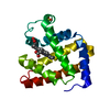
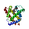
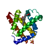
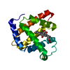
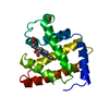
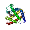
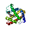
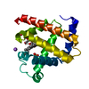
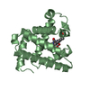
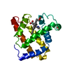
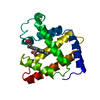
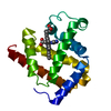
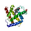
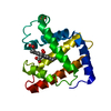
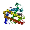
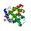
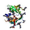
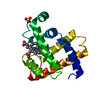
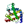
| Title | CRYSTAL STRUCTURE OF OXY SPERM WHALE MYOGLOBIN MUTANT Y(B10)Q(E7)R(E10) | ||||||
 Components Components | MYOGLOBIN | ||||||
 Keywords Keywords | OXYGEN STORAGE/TRANSPORT / MYOGLOBIN / HEME / TRIPLE MUTANT / OXYGEN STORAGE-TRANSPORT COMPLEX | ||||||
| Function / homology |  Function and homology information Function and homology informationOxidoreductases; Acting on other nitrogenous compounds as donors / nitrite reductase activity / sarcoplasm / Oxidoreductases; Acting on a peroxide as acceptor; Peroxidases / removal of superoxide radicals / oxygen carrier activity / peroxidase activity / oxygen binding / heme binding / extracellular exosome / metal ion binding Similarity search - Function | ||||||
| Biological species |  | ||||||
| Method |  X-RAY DIFFRACTION / Resolution: 1.7 Å X-RAY DIFFRACTION / Resolution: 1.7 Å | ||||||
 Authors Authors | Brunori, M. / Cutruzzola, F. / Savino, C. / Travaglini-Allocatelli, C. / Vallone, B. / Gibson, Q.H. | ||||||
 Citation Citation | Journal: Biophys.J. / Year: 1999 Title: Structural dynamics of ligand diffusion in the protein matrix: A study on a new myoglobin mutant Y(B10) Q(E7) R(E10). Authors: Brunori, M. / Cutruzzola, F. / Savino, C. / Travaglini-Allocatelli, C. / Vallone, B. / Gibson, Q.H. #1: Journal: TRENDS BIOCHEM.SCI. / Year: 1999Title: Does Picosecond Protein Dynamics Have a Survival Value? Authors: Brunori, M. / Cutruzzola, F. / Savino, C. / Travaglini-Allocatelli, C. / Vallone, B. / Gibson, Q.H. #2: Journal: Proc.Natl.Acad.Sci.USA / Year: 2000Title: The Role of Cavities in Protein Dynamics: Crystal Structure of a Photolytic Intermediate of a Mutant Myoglobin Authors: Brunori, M. / Vallone, B. / Cutruzzola, F. / Travaglini-Allocatelli, C. / Berendzen, J. / Chu, K. / Sweet, R.M. / Schlichting, I. | ||||||
| History |
|
- Structure visualization
| Structure viewer | Molecule: MolmilJmol/JSmol |
|---|
- Downloads & links
-Download
| PDBx/mmCIF format | 1f65.cif.gz | 50.2 KB | Display | PDBx/mmCIF format |
|---|---|---|---|---|
| PDB format | pdb1f65.ent.gz | 34.9 KB | Display | PDB format |
| PDBx/mmJSON format | 1f65.json.gz | Tree view | PDBx/mmJSON format | |
| Others |  Other downloads Other downloads |
-Validation report
| Arichive directory | https://data.pdbj.org/pub/pdb/validation_reports/f6/1f65ftp://data.pdbj.org/pub/pdb/validation_reports/f6/1f65 | HTTPS FTP |
|---|
-Related structure data


-Links
 PDBj
PDBj


- Assembly
| Deposited unit | 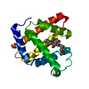
| ||||||||
|---|---|---|---|---|---|---|---|---|---|
| 1 |
| ||||||||
| Unit cell |
|
-Components
| #1: Protein | Mass: 17461.250 Da / Num. of mol.: 1 / Mutation: L29Y, H64Q, T67R Source method: isolated from a genetically manipulated source Source: (gene. exp.)  | ||||||||
|---|---|---|---|---|---|---|---|---|---|
| #2: Chemical |   Mass: 96.063 Da / Num. of mol.: 2 / Source method: obtained synthetically / Formula: SO4 Mass: 96.063 Da / Num. of mol.: 2 / Source method: obtained synthetically / Formula: SO4#3: Chemical | ChemComp-HEM / |   Mass: 616.487 Da / Num. of mol.: 1 / Source method: obtained synthetically / Formula: C34H32FeN4O4 Mass: 616.487 Da / Num. of mol.: 1 / Source method: obtained synthetically / Formula: C34H32FeN4O4#4: Chemical | ChemComp-OXY / |   Mass: 31.999 Da / Num. of mol.: 1 / Source method: obtained synthetically / Formula: O2 Mass: 31.999 Da / Num. of mol.: 1 / Source method: obtained synthetically / Formula: O2#5: Water | ChemComp-HOH / |  Mass: 18.015 Da / Num. of mol.: 167 / Source method: isolated from a natural source / Formula: H2O Mass: 18.015 Da / Num. of mol.: 167 / Source method: isolated from a natural source / Formula: H2OCompound details | Triple Mb mutant designed to mimick the properties of Ascaris suum Hb. We observed a H-bonding ...Triple Mb mutant designed to mimick the properties of Ascaris suum Hb. We observed a H-bonding pattern to heme bound O2 which is virtually identical to the one found in A. suum Hb. | |
-Experimental details
-Experiment
| Experiment | Method: X-RAY DIFFRACTION / Number of used crystals: 1 |
|---|
- Sample preparation
| Crystal | Density Matthews: 3.05 Å3/Da / Density % sol: 59.68 % | ||||||||||||||||||||
|---|---|---|---|---|---|---|---|---|---|---|---|---|---|---|---|---|---|---|---|---|---|
| Crystal grow | Temperature: 293 K / Method: vapor diffusion, hanging drop / pH: 8.6 Details: 2.7 M Ammonium sulphate, 20 mM Tris-Cl, 1mM EDTA reduced with sodium dithionate and subsequently derivatised with dioxigen, pH 8.6, VAPOR DIFFUSION, HANGING DROP, temperature 293K | ||||||||||||||||||||
| Crystal grow | *PLUS Temperature: 21 ℃ / pH: 8.7 / Method: vapor diffusionDetails: drop consists of equal volume of protein and reservoir solutions | ||||||||||||||||||||
| Components of the solutions | *PLUS
|
-Data collection
| Diffraction | Mean temperature: 120 K |
|---|---|
| Diffraction source | Source: ROTATING ANODE / Type: RIGAKU RU200 / Wavelength: 1.5418 |
| Detector | Type: RIGAKU RAXIS II / Detector: IMAGE PLATE / Date: May 1, 1998 |
| Radiation | Protocol: SINGLE WAVELENGTH / Monochromatic (M) / Laue (L): M / Scattering type: x-ray |
| Radiation wavelength | Wavelength: 1.5418 Å / Relative weight: 1 |
| Reflection | Resolution: 1.7→14.8 Å / Num. all: 26633 / Num. obs: 26622 / % possible obs: 95.1 % / Observed criterion σ(F): 2 / Observed criterion σ(I): 2 / Redundancy: 7 % / Biso Wilson estimate: 17.5 Å2 / Rmerge(I) obs: 0.083 / Net I/σ(I): 15.6 |
| Reflection shell | Resolution: 1.7→1.8 Å / Redundancy: 8 % / Rmerge(I) obs: 0.143 / Num. unique all: 2511 / % possible all: 90.7 |
| Reflection shell | *PLUS % possible obs: 90.7 % |
- Processing
| Software |
| |||||||||||||||||||||||||
|---|---|---|---|---|---|---|---|---|---|---|---|---|---|---|---|---|---|---|---|---|---|---|---|---|---|---|
| Refinement | Resolution: 1.7→14.8 Å / σ(F): 2 / σ(I): 2 / Stereochemistry target values: PROTIN
| |||||||||||||||||||||||||
| Refinement step | Cycle: LAST / Resolution: 1.7→14.8 Å
| |||||||||||||||||||||||||
| Refine LS restraints |
| |||||||||||||||||||||||||
| Software | *PLUS Name: REFMAC / Classification: refinement | |||||||||||||||||||||||||
| Refine LS restraints | *PLUS
|
