 Movie
Movie Controller

[English] 日本語
 Yorodumi
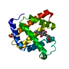
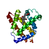
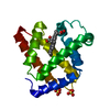
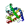
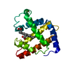
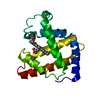
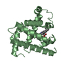
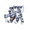
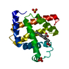
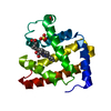
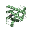
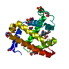
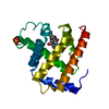
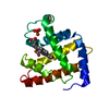
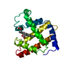
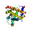
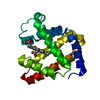
- PDB-1f63: CRYSTAL STRUCTURE OF DEOXY SPERM WHALE MYOGLOBIN MUTANT Y(B10)Q(E... -
+ Open data

- Basic information
| Entry | Database: PDB / ID: 1f63 | ||||||
|---|---|---|---|---|---|---|---|
| Title | CRYSTAL STRUCTURE OF DEOXY SPERM WHALE MYOGLOBIN MUTANT Y(B10)Q(E7)R(E10) | ||||||
 Components Components | MYOGLOBIN | ||||||
 Keywords Keywords | OXYGEN STORAGE/TRANSPORT / myoglobin / heme / triple mutant / OXYGEN STORAGE-TRANSPORT COMPLEX | ||||||
| Function / homology |  Function and homology information Function and homology informationOxidoreductases; Acting on other nitrogenous compounds as donors / nitrite reductase activity / sarcoplasm / Oxidoreductases; Acting on a peroxide as acceptor; Peroxidases / removal of superoxide radicals / oxygen carrier activity / peroxidase activity / oxygen binding / heme binding / extracellular exosome / metal ion binding Similarity search - Function | ||||||
| Biological species |  | ||||||
| Method |  X-RAY DIFFRACTION / Resolution: 1.8 Å X-RAY DIFFRACTION / Resolution: 1.8 Å | ||||||
 Authors Authors | Brunori, M. / Cutruzzola, F. / Savino, C. / Travaglini-Allocatelli, C. / Vallone, B. / Gibson, Q.H. | ||||||
 Citation Citation | Journal: Biophys.J. / Year: 1999 Title: Structural dynamics of ligand diffusion in the protein matrix: A study on a new myoglobin mutant Y(B10) Q(E7) R(E10). Authors: Brunori, M. / Cutruzzola, F. / Savino, C. / Travaglini-Allocatelli, C. / Vallone, B. / Gibson, Q.H. #1: Journal: TRENDS BIOCHEM.SCI. / Year: 1999Title: Does Picosecond Protein Dynamics Have a Survival Value? Authors: Brunori, M. / Cutruzzola, F. / Savino, C. / Travaglini-Allocatelli, C. / Vallone, B. / Gibson, Q.H. #2: Journal: Proc.Natl.Acad.Sci.USA / Year: 2000Title: The Role of Cavities in Protein Dynamics: Crystal Structure of a Photolytic Intermediate of a Mutant Myoglobin Authors: Brunori, M. / Vallone, B. / Cutruzzola, F. / Travaglini-Allocatelli, C. / Berendzen, J. / Chu, K. / Sweet, R.M. / Schlichting, I. | ||||||
| History |
|
- Structure visualization
| Structure viewer | Molecule: MolmilJmol/JSmol |
|---|
- Downloads & links
-Download
| PDBx/mmCIF format | 1f63.cif.gz | 47.6 KB | Display | PDBx/mmCIF format |
|---|---|---|---|---|
| PDB format | pdb1f63.ent.gz | 33.4 KB | Display | PDB format |
| PDBx/mmJSON format | 1f63.json.gz | Tree view | PDBx/mmJSON format | |
| Others |  Other downloads Other downloads |
-Validation report
| Arichive directory | https://data.pdbj.org/pub/pdb/validation_reports/f6/1f63ftp://data.pdbj.org/pub/pdb/validation_reports/f6/1f63 | HTTPS FTP |
|---|
-Related structure data


-Links
 PDBj
PDBj


- Assembly
| Deposited unit | 
| ||||||||
|---|---|---|---|---|---|---|---|---|---|
| 1 |
| ||||||||
| Unit cell |
|
-Components
| #1: Protein | Mass: 17461.250 Da / Num. of mol.: 1 / Mutation: L29Y, H64Q, T67R Source method: isolated from a genetically manipulated source Source: (gene. exp.)  |
|---|---|
| #2: Chemical | ChemComp-SO4 /   Mass: 96.063 Da / Num. of mol.: 1 / Source method: obtained synthetically / Formula: SO4 Mass: 96.063 Da / Num. of mol.: 1 / Source method: obtained synthetically / Formula: SO4 |
| #3: Chemical | ChemComp-HEM /   Mass: 616.487 Da / Num. of mol.: 1 / Source method: obtained synthetically / Formula: C34H32FeN4O4 Mass: 616.487 Da / Num. of mol.: 1 / Source method: obtained synthetically / Formula: C34H32FeN4O4 |
| #4: Water | ChemComp-HOH /  Mass: 18.015 Da / Num. of mol.: 90 / Source method: isolated from a natural source / Formula: H2O Mass: 18.015 Da / Num. of mol.: 90 / Source method: isolated from a natural source / Formula: H2O |
| Compound details | Triple Mb mutant designed to mimick the properties of Ascaris suum Hb. The amino acid in the heme ...Triple Mb mutant designed to mimick the properties of Ascaris suum Hb. The amino acid in the heme distal site do not allow ligand binding to the heme without movement of the mutated Y(B10) and Q(E7) |
-Experimental details
-Experiment
| Experiment | Method: X-RAY DIFFRACTION / Number of used crystals: 1 |
|---|
- Sample preparation
| Crystal | Density Matthews: 3.18 Å3/Da / Density % sol: 61.35 % | |||||||||||||||||||||||||
|---|---|---|---|---|---|---|---|---|---|---|---|---|---|---|---|---|---|---|---|---|---|---|---|---|---|---|
| Crystal grow | Temperature: 293 K / Method: vapor diffusion, hanging drop / pH: 8.7 Details: 2.7 M Ammonium sulphate, 20 mM Tris-Cl, 1 mM EDTA, pH 8.7, VAPOR DIFFUSION, HANGING DROP, temperature 293K | |||||||||||||||||||||||||
| Crystal grow | *PLUS Temperature: 21 ℃ / Method: vapor diffusionDetails: drop consists of equal volume of protein and reservoir solutions | |||||||||||||||||||||||||
| Components of the solutions | *PLUS
|
-Data collection
| Diffraction | Mean temperature: 293 K |
|---|---|
| Diffraction source | Source: ROTATING ANODE / Type: RIGAKU RU200 / Wavelength: 1.5418 |
| Detector | Type: RIGAKU RAXIS II / Detector: IMAGE PLATE / Date: Sep 1, 1998 |
| Radiation | Protocol: SINGLE WAVELENGTH / Monochromatic (M) / Laue (L): M / Scattering type: x-ray |
| Radiation wavelength | Wavelength: 1.5418 Å / Relative weight: 1 |
| Reflection | Resolution: 1.8→15 Å / Num. all: 60982 / Num. obs: 19613 / % possible obs: 94.2 % / Observed criterion σ(F): 2 / Observed criterion σ(I): 2 / Redundancy: 3.1 % / Biso Wilson estimate: 18.5 Å2 / Rmerge(I) obs: 0.095 / Net I/σ(I): 13 |
| Reflection shell | Resolution: 1.8→15 Å / Redundancy: 2 % / Rmerge(I) obs: 0.249 / Num. unique all: 1079 / % possible all: 89.3 |
| Reflection shell | *PLUS % possible obs: 89.3 % |
- Processing
| Software |
| |||||||||||||||||||||||||
|---|---|---|---|---|---|---|---|---|---|---|---|---|---|---|---|---|---|---|---|---|---|---|---|---|---|---|
| Refinement | Resolution: 1.8→15 Å / σ(F): 3 / σ(I): 3 / Stereochemistry target values: PROTIN
| |||||||||||||||||||||||||
| Refinement step | Cycle: LAST / Resolution: 1.8→15 Å
| |||||||||||||||||||||||||
| Refine LS restraints |
| |||||||||||||||||||||||||
| Software | *PLUS Name: REFMAC / Classification: refinement | |||||||||||||||||||||||||
| Refine LS restraints | *PLUS
|
