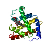
 Movie
Movie Controller


+ Open data

- Basic information
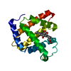
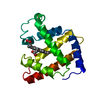
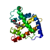
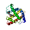
| Entry | Database: PDB / ID: 1dxc | ||||||
|---|---|---|---|---|---|---|---|
| Title | CO complex of Myoglobin Mb-YQR at 100K | ||||||
 Components Components | MYOGLOBIN | ||||||
 Keywords Keywords | OXYGEN STORAGE / CO COMPLEX / RESPIRATORY PROTEIN | ||||||
| Function / homology |  Function and homology information Function and homology informationOxidoreductases; Acting on other nitrogenous compounds as donors / nitrite reductase activity / sarcoplasm / Oxidoreductases; Acting on a peroxide as acceptor; Peroxidases / removal of superoxide radicals / oxygen carrier activity / peroxidase activity / oxygen binding / heme binding / extracellular exosome / metal ion binding Similarity search - Function | ||||||
| Biological species |  | ||||||
| Method |  X-RAY DIFFRACTION / SYNCHROTRON / MOLECULAR REPLACEMENT / Resolution: 1.4 Å X-RAY DIFFRACTION / SYNCHROTRON / MOLECULAR REPLACEMENT / Resolution: 1.4 Å | ||||||
 Authors Authors | Brunori, M. / Vallone, B. / Cutruzzola, F. / Travaglini-Allocatelli, C. / Berendzen, J. / Chu, K. / Sweet, R.M. / Schlichting, I. | ||||||
 Citation Citation | Journal: Proc.Natl.Acad.Sci.USA / Year: 2000 Title: The Role of Cavities in Protein Dynamics: Crystal Structure of a Novel Photolytic Intermediate of Myoglobin Authors: Brunori, M. / Vallone, B. / Cutruzzola, F. / Travaglini-Allocatelli, C. / Berendzen, J. / Chu, K. / Sweet, R.M. / Schlichting, I. #1: Journal: Biophys.J. / Year: 1999Title: Structural Dynamics of Liganddiffusion in the Protein Matrix: A Study on a New Myoglobin Mutant Y (B10) Q(E7) R(E10) Authors: Brunori, M. / Cutruzzola, F. / Savino, C. / Travaglini-Allocatelli, C. / Vallone, B. / Gibson, Q.H. #2: Journal: Nature / Year: 1994Title: Crystal Structure of Photolysed Carbonmonoxy-Myoglobin Authors: Schlichting, I. / Berendzen, J. / Juniorsweet, R.M.G.N.P. #3: Journal: Proteins: Struct.,Funct., Genet. / Year: 1990 Title: Crystal Structure of Myoglobin from a Synthetic Gene Authors: Juniorarduini, R.M.G.N.P. / Springer, B.A. / Sligar, S.G. #4: Journal: Proc.Natl.Acad.Sci.USA / Year: 1987 Title: High-Level Expression of Sperm Whale Myoglobin in Escherichia Coli Authors: Springer, B.A. / Sligar, S.G. | ||||||
| History |
|
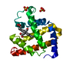
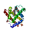
- Structure visualization
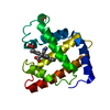
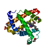
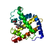
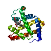
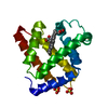
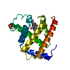
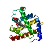
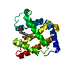
| Structure viewer | Molecule: MolmilJmol/JSmol |
|---|
- Downloads & links
-Download
| PDBx/mmCIF format | 1dxc.cif.gz | 90 KB | Display | PDBx/mmCIF format |
|---|---|---|---|---|
| PDB format | pdb1dxc.ent.gz | 67.8 KB | Display | PDB format |
| PDBx/mmJSON format | 1dxc.json.gz | Tree view | PDBx/mmJSON format | |
| Others |  Other downloads Other downloads |
-Validation report
| Arichive directory | https://data.pdbj.org/pub/pdb/validation_reports/dx/1dxcftp://data.pdbj.org/pub/pdb/validation_reports/dx/1dxc | HTTPS FTP |
|---|
-Related structure data


-Links
 PDBj
PDBj


- Assembly
| Deposited unit | 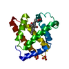
| ||||||||
|---|---|---|---|---|---|---|---|---|---|
| 1 |
| ||||||||
| Unit cell |
| ||||||||
| Components on special symmetry positions |
| ||||||||
| Details | IN THIS SPACE GROUP THERE IS A STRONG 3 -FOLD CRYSTAL PACKINGOF THE MYOGLOBIN MOLECULE INVOLVING SYMMETRY OPERATIONS,(X,Y,Z), (1.0-X+ Y,1.0-X,Z), AND (1.0-Y,X-Y,Z). THE SO4MOLECULE 315A SITS CLOSE TO THE 3-FOLD AXIS. |
-Components
| #1: Protein | Mass: 17461.250 Da / Num. of mol.: 1 / Mutation: YES Source method: isolated from a genetically manipulated source Details: HEME BOUND TO HIS-93, CO BOUND TO HEME IRON / Source: (gene. exp.)  | ||||
|---|---|---|---|---|---|
| #2: Chemical | ChemComp-HEM /   Mass: 616.487 Da / Num. of mol.: 1 / Source method: obtained synthetically / Formula: C34H32FeN4O4 Mass: 616.487 Da / Num. of mol.: 1 / Source method: obtained synthetically / Formula: C34H32FeN4O4 | ||||
| #3: Chemical | ChemComp-CMO /   Mass: 28.010 Da / Num. of mol.: 1 / Source method: obtained synthetically / Formula: CO Mass: 28.010 Da / Num. of mol.: 1 / Source method: obtained synthetically / Formula: CO | ||||
| #4: Chemical |   Mass: 96.063 Da / Num. of mol.: 3 / Source method: obtained synthetically / Formula: SO4 Mass: 96.063 Da / Num. of mol.: 3 / Source method: obtained synthetically / Formula: SO4#5: Water | ChemComp-HOH / |  Mass: 18.015 Da / Num. of mol.: 258 / Source method: isolated from a natural source / Formula: H2O Mass: 18.015 Da / Num. of mol.: 258 / Source method: isolated from a natural source / Formula: H2OCompound details | CO IS ATTACHED TO HEME IRON ATOM WHICH IS COORDINATE | |
-Experimental details
-Experiment
| Experiment | Method: X-RAY DIFFRACTION / Number of used crystals: 1 |
|---|
- Sample preparation
| Crystal | Density Matthews: 3.08 Å3/Da / Density % sol: 60.02 % | |||||||||||||||||||||||||
|---|---|---|---|---|---|---|---|---|---|---|---|---|---|---|---|---|---|---|---|---|---|---|---|---|---|---|
| Crystal grow | pH: 9 Details: PROTEIN WAS CRYSTALLIZED FROM 3.6 M AMMONIUM SULFATE.CRYSTALS WERE SOAKED FOR C.A. 1 HOUR IN A THOROUGHLY DEGASSED AND CO SATURATED CRYOPROTECTANT SOLUTION MADE BY ADDITION OF 100 MG ...Details: PROTEIN WAS CRYSTALLIZED FROM 3.6 M AMMONIUM SULFATE.CRYSTALS WERE SOAKED FOR C.A. 1 HOUR IN A THOROUGHLY DEGASSED AND CO SATURATED CRYOPROTECTANT SOLUTION MADE BY ADDITION OF 100 MG XYLITOL, 100 MG GLUCOSE AND 8 MG NA-DITHIONATE TO 1 ML OF 70% SATURATED AMMONIUM SULFATE WITH 50 MM TRIS.HCL PH 9.0. DISTINCT COLOR CHANGES INDICATED THE FORMATION OF CO-MB FROM MET-MB. | |||||||||||||||||||||||||
| Crystal grow | *PLUS Temperature: 21 ℃ / pH: 8.7 / Method: vapor diffusion | |||||||||||||||||||||||||
| Components of the solutions | *PLUS
|
-Data collection
| Diffraction | Mean temperature: 100 K |
|---|---|
| Diffraction source | Source: SYNCHROTRON / Site: NSLS  / Beamline: X12C / Wavelength: 0.91 / Beamline: X12C / Wavelength: 0.91 |
| Detector | Type: PRINCETON SCIENTIFIC / Detector: CCD / Date: May 15, 1998 / Details: MIRRORS |
| Radiation | Monochromator: YES / Protocol: SINGLE WAVELENGTH / Monochromatic (M) / Laue (L): M / Scattering type: x-ray |
| Radiation wavelength | Wavelength: 0.91 Å / Relative weight: 1 |
| Reflection | Resolution: 1.4→19.6 Å / Num. obs: 40518 / % possible obs: 96.5 % / Observed criterion σ(I): 0 / Redundancy: 3.5 % / Biso Wilson estimate: 15.2 Å2 / Rsym value: 0.036 / Net I/σ(I): 19.8 |
| Reflection shell | Resolution: 1.4→1.5 Å / Redundancy: 2.1 % / Mean I/σ(I) obs: 3.6 / Rsym value: 0.125 / % possible all: 91.4 |
| Reflection | *PLUS Lowest resolution: 19.6 Å / Num. obs: 40518 / % possible obs: 96.5 % / Num. measured all: 143218 / Rmerge(I) obs: 0.036 |
| Reflection shell | *PLUS % possible obs: 91.4 % / Num. unique obs: 7138 / Num. measured obs: 15373 / Rmerge(I) obs: 0.125 / Mean I/σ(I) obs: 5.8 |
- Processing
| Software |
| |||||||||||||||||||||||||||||||||||||||||||||||||||||||||||||||
|---|---|---|---|---|---|---|---|---|---|---|---|---|---|---|---|---|---|---|---|---|---|---|---|---|---|---|---|---|---|---|---|---|---|---|---|---|---|---|---|---|---|---|---|---|---|---|---|---|---|---|---|---|---|---|---|---|---|---|---|---|---|---|---|---|
| Refinement | Method to determine structure: MOLECULAR REPLACEMENT / Resolution: 1.4→19.6 Å / Cross valid method: THROUGHOUT / σ(F): 0
| |||||||||||||||||||||||||||||||||||||||||||||||||||||||||||||||
| Refinement step | Cycle: LAST / Resolution: 1.4→19.6 Å
| |||||||||||||||||||||||||||||||||||||||||||||||||||||||||||||||
| Refine LS restraints |
| |||||||||||||||||||||||||||||||||||||||||||||||||||||||||||||||
| Software | *PLUS Name: REFMAC / Classification: refinement | |||||||||||||||||||||||||||||||||||||||||||||||||||||||||||||||
| Refinement | *PLUS Lowest resolution: 19.6 Å / Rfactor obs: 0.129 / Rfactor Rfree: 0.153 / Rfactor Rwork: 0.12845 | |||||||||||||||||||||||||||||||||||||||||||||||||||||||||||||||
| Solvent computation | *PLUS | |||||||||||||||||||||||||||||||||||||||||||||||||||||||||||||||
| Displacement parameters | *PLUS | |||||||||||||||||||||||||||||||||||||||||||||||||||||||||||||||
| Refine LS restraints | *PLUS Type: p_bond_d / Dev ideal: 0.014 |
