 Movie
Movie Controller

[English] 日本語
 Yorodumi
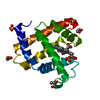
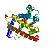
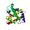
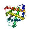
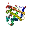
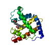
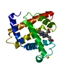
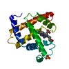
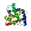
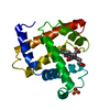
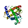
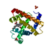
- PDB-1mbo: Structure and refinement of oxymyoglobin at 1.6 angstroms resolution -
+ Open data

- Basic information
| Entry | Database: PDB / ID: 1mbo | |||||||||
|---|---|---|---|---|---|---|---|---|---|---|
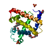
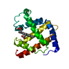
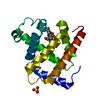
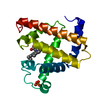
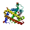
| Title | Structure and refinement of oxymyoglobin at 1.6 angstroms resolution | |||||||||
 Components Components | MYOGLOBIN | |||||||||
 Keywords Keywords | OXYGEN STORAGE | |||||||||
| Function / homology |  Function and homology information Function and homology informationnitrite reductase activity / Oxidoreductases; Acting on other nitrogenous compounds as donors / sarcoplasm / Oxidoreductases; Acting on a peroxide as acceptor; Peroxidases / removal of superoxide radicals / oxygen carrier activity / peroxidase activity / oxygen binding / heme binding / extracellular exosome / metal ion binding Similarity search - Function | |||||||||
| Biological species |  | |||||||||
| Method |  X-RAY DIFFRACTION / Resolution: 1.6 Å X-RAY DIFFRACTION / Resolution: 1.6 Å | |||||||||
 Authors Authors | Phillips, S.E.V. | |||||||||
 Citation Citation | Journal: J.Mol.Biol. / Year: 1980 Title: Structure and refinement of oxymyoglobin at 1.6 A resolution. Authors: Phillips, S.E. #1: Journal: Nature / Year: 1981Title: Neutron Diffraction Reveals Oxygen-Histidine Hydrogen Bond in Oxymyoglobin Authors: Phillips, S.E.V. / Schoenborn, B.P. #3: Journal: Acta Crystallogr.,Sect.A (Supplement) / Year: 1978Title: The Structure of Oxy-Myoglobin Authors: Phillips, S.E.V. | |||||||||
| History |
|
- Structure visualization
| Structure viewer | Molecule: MolmilJmol/JSmol |
|---|
- Downloads & links
-Download
| PDBx/mmCIF format | 1mbo.cif.gz | 54.9 KB | Display | PDBx/mmCIF format |
|---|---|---|---|---|
| PDB format | pdb1mbo.ent.gz | 36.2 KB | Display | PDB format |
| PDBx/mmJSON format | 1mbo.json.gz | Tree view | PDBx/mmJSON format | |
| Others |  Other downloads Other downloads |
-Validation report
| Arichive directory | https://data.pdbj.org/pub/pdb/validation_reports/mb/1mboftp://data.pdbj.org/pub/pdb/validation_reports/mb/1mbo | HTTPS FTP |
|---|
-Related structure data
| Similar structure data |
|---|


-Links
 PDBj
PDBj


- Assembly
| Deposited unit | 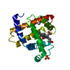
| ||||||||
|---|---|---|---|---|---|---|---|---|---|
| 1 |
| ||||||||
| Unit cell |
| ||||||||
| Atom site foot note | 1: INSPECTION OF MAPS AT VARIOUS STAGES OF REFINEMENT INDICATED THAT SEVERAL SIDE-CHAINS AND THE C- AND N-TERMINAL RESIDUES WERE DISORDERED TO SOME DEGREE. IN MOST CASES, ONE CONFORMATION WAS VERY ...1: INSPECTION OF MAPS AT VARIOUS STAGES OF REFINEMENT INDICATED THAT SEVERAL SIDE-CHAINS AND THE C- AND N-TERMINAL RESIDUES WERE DISORDERED TO SOME DEGREE. IN MOST CASES, ONE CONFORMATION WAS VERY MUCH STRONGER THAN THE OTHER, BUT FOUR SIDE-CHAINS WERE INCLUDED IN THE MODEL WITH TWO ALTERNATE CONFORMATIONS (VAL 13, LEU 86, LEU 89, GLN 128). 2: HOH 305 IS CLOSE TO ATOM O1 OF HEM 546. SEE PAPER CITED AS JRNL REFERENCE ABOVE. |
-Components
| #1: Protein | Mass: 17234.951 Da / Num. of mol.: 1 Source method: isolated from a genetically manipulated source Source: (gene. exp.) |
|---|---|
| #2: Chemical | ChemComp-SO4 /   Mass: 96.063 Da / Num. of mol.: 1 / Source method: obtained synthetically / Formula: SO4 Mass: 96.063 Da / Num. of mol.: 1 / Source method: obtained synthetically / Formula: SO4 |
| #3: Chemical | ChemComp-HEM /   Mass: 616.487 Da / Num. of mol.: 1 / Source method: obtained synthetically / Formula: C34H32FeN4O4 Mass: 616.487 Da / Num. of mol.: 1 / Source method: obtained synthetically / Formula: C34H32FeN4O4 |
| #4: Chemical | ChemComp-OXY /   Mass: 31.999 Da / Num. of mol.: 1 / Source method: obtained synthetically / Formula: O2 Mass: 31.999 Da / Num. of mol.: 1 / Source method: obtained synthetically / Formula: O2 |
| #5: Water | ChemComp-HOH /  Mass: 18.015 Da / Num. of mol.: 334 / Source method: isolated from a natural source / Formula: H2O Mass: 18.015 Da / Num. of mol.: 334 / Source method: isolated from a natural source / Formula: H2O |
-Experimental details
-Experiment
| Experiment | Method: X-RAY DIFFRACTION |
|---|
- Sample preparation
| Crystal | Density Matthews: 1.95 Å3/Da / Density % sol: 37.03 % |
|---|---|
| Crystal grow | *PLUS Temperature: 4 ℃ / pH: 8.4 / Method: unknown |
-Data collection
| Radiation | Protocol: SINGLE WAVELENGTH / Monochromatic (M) / Laue (L): M / Scattering type: x-ray |
|---|---|
| Radiation wavelength | Relative weight: 1 |
| Reflection | *PLUS Highest resolution: 1.6 Å / Lowest resolution: 20 Å / Num. obs: 18494 / Num. measured all: 53607 / Rmerge(I) obs: 0.065 |
- Processing
| Software | Name: CONSTRAINED / Version: RECIPROCAL-SPACE LEAST-SQUARES / Classification: refinement | ||||||||||||
|---|---|---|---|---|---|---|---|---|---|---|---|---|---|
| Refinement | Highest resolution: 1.6 Å | ||||||||||||
| Refinement step | Cycle: LAST / Highest resolution: 1.6 Å
| ||||||||||||
| Refinement | *PLUS Num. reflection obs: 18494 / σ(F): 3 / Rfactor all: 0.159 / Rfactor obs: 0.153 | ||||||||||||
| Solvent computation | *PLUS | ||||||||||||
| Displacement parameters | *PLUS |
