 Movie
Movie Controller

[English] 日本語
 Yorodumi
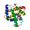
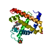
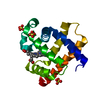
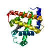
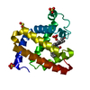
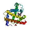
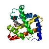
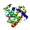
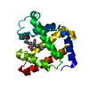
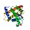
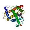
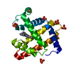
- PDB-1bzp: ATOMIC RESOLUTION CRYSTAL STRUCTURE ANALYSIS OF NATIVE DEOXY AND ... -
+ Open data

- Basic information
| Entry | Database: PDB / ID: 1bzp | ||||||
|---|---|---|---|---|---|---|---|
| Title | ATOMIC RESOLUTION CRYSTAL STRUCTURE ANALYSIS OF NATIVE DEOXY AND CO MYOGLOBIN FROM SPERM WHALE AT ROOM TEMPERATURE | ||||||
 Components Components | PROTEIN (MYOGLOBIN) | ||||||
 Keywords Keywords | OXYGEN STORAGE / DEOXY MYOGLOBIN / ATOMIC RESOLUTION | ||||||
| Function / homology |  Function and homology information Function and homology informationOxidoreductases; Acting on other nitrogenous compounds as donors / nitrite reductase activity / sarcoplasm / Oxidoreductases; Acting on a peroxide as acceptor; Peroxidases / removal of superoxide radicals / oxygen carrier activity / peroxidase activity / oxygen binding / heme binding / extracellular exosome / metal ion binding Similarity search - Function | ||||||
| Biological species |  | ||||||
| Method |  X-RAY DIFFRACTION / SYNCHROTRON / MOLECULAR REPLACEMENT / Resolution: 1.15 Å X-RAY DIFFRACTION / SYNCHROTRON / MOLECULAR REPLACEMENT / Resolution: 1.15 Å | ||||||
 Authors Authors | Kachalova, G.S. / Popov, A.N. / Bartunik, H.D. | ||||||
 Citation Citation | Journal: Science / Year: 1999 Title: A steric mechanism for inhibition of CO binding to heme proteins. Authors: Kachalova, G.S. / Popov, A.N. / Bartunik, H.D. | ||||||
| History |
|
- Structure visualization
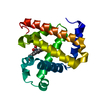
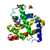
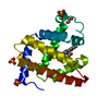
| Structure viewer | Molecule: MolmilJmol/JSmol |
|---|
- Downloads & links
-Download
| PDBx/mmCIF format | 1bzp.cif.gz | 90.8 KB | Display | PDBx/mmCIF format |
|---|---|---|---|---|
| PDB format | pdb1bzp.ent.gz | 68.3 KB | Display | PDB format |
| PDBx/mmJSON format | 1bzp.json.gz | Tree view | PDBx/mmJSON format | |
| Others |  Other downloads Other downloads |
-Validation report
| Arichive directory | https://data.pdbj.org/pub/pdb/validation_reports/bz/1bzpftp://data.pdbj.org/pub/pdb/validation_reports/bz/1bzp | HTTPS FTP |
|---|
-Related structure data
| Related structure data | 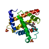 1bz6C 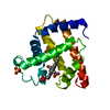 1bzrC 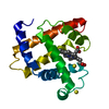 4mbnS S: Starting model for refinement C: citing same article ( |
|---|---|
| Similar structure data |

-Links
 PDBj
PDBj


- Assembly
| Deposited unit | 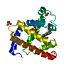
| ||||||||
|---|---|---|---|---|---|---|---|---|---|
| 1 |
| ||||||||
| Unit cell |
|
-Components
| #1: Protein | Mass: 17234.951 Da / Num. of mol.: 1 / Source method: isolated from a natural source / Source: (natural) | ||||
|---|---|---|---|---|---|
| #2: Chemical |   Mass: 96.063 Da / Num. of mol.: 3 / Source method: obtained synthetically / Formula: SO4 Mass: 96.063 Da / Num. of mol.: 3 / Source method: obtained synthetically / Formula: SO4#3: Chemical | ChemComp-HEM / |   Mass: 616.487 Da / Num. of mol.: 1 / Source method: obtained synthetically / Formula: C34H32FeN4O4 Mass: 616.487 Da / Num. of mol.: 1 / Source method: obtained synthetically / Formula: C34H32FeN4O4#4: Water | ChemComp-HOH / |  Mass: 18.015 Da / Num. of mol.: 226 / Source method: isolated from a natural source / Formula: H2O Mass: 18.015 Da / Num. of mol.: 226 / Source method: isolated from a natural source / Formula: H2O |
-Experimental details
-Experiment
| Experiment | Method: X-RAY DIFFRACTION / Number of used crystals: 1 |
|---|
- Sample preparation
| Crystal | Density Matthews: 1.97 Å3/Da / Density % sol: 37.53 % |
|---|---|
| Crystal grow | pH: 5.9 Details: PROTEIN WAS CRYSTALLIZED FROM AMMONIUM SULPHATE, 0.1 M POTASSIUM PHOSPHATE., pH 5.9 |
| Crystal grow | *PLUS Method: unknown |
-Data collection
| Diffraction | Mean temperature: 287 K |
|---|---|
| Diffraction source | Source: SYNCHROTRON / Site: MPG/DESY, HAMBURG  / Beamline: BW6 / Wavelength: 0.9 / Beamline: BW6 / Wavelength: 0.9 |
| Detector | Type: MAR scanner 300 mm plate / Detector: IMAGE PLATE / Date: Apr 1, 1997 / Details: MIRRORS |
| Radiation | Monochromator: SI(111) / Protocol: SINGLE WAVELENGTH / Monochromatic (M) / Laue (L): M / Scattering type: x-ray |
| Radiation wavelength | Wavelength: 0.9 Å / Relative weight: 1 |
| Reflection | Resolution: 1.15→20 Å / Num. obs: 41207 / % possible obs: 85.9 % / Redundancy: 4.2 % / Rmerge(I) obs: 0.062 / Net I/σ(I): 19.9 |
| Reflection shell | Resolution: 1.15→1.2 Å / Redundancy: 2.7 % / Rmerge(I) obs: 0.31 / Mean I/σ(I) obs: 2.7 / % possible all: 66.1 |
| Reflection | *PLUS Num. measured all: 175253 |
| Reflection shell | *PLUS % possible obs: 66.1 % |
- Processing
| Software |
| |||||||||||||||||||||||||||||||||
|---|---|---|---|---|---|---|---|---|---|---|---|---|---|---|---|---|---|---|---|---|---|---|---|---|---|---|---|---|---|---|---|---|---|---|
| Refinement | Method to determine structure: MOLECULAR REPLACEMENT Starting model: 4MBN Resolution: 1.15→12 Å / Num. parameters: 14105 / Num. restraintsaints: 18174 / σ(F): 0 StereochEM target val spec case: HEME - PARAMETERS BASED ON CSD Stereochemistry target values: ENGH AND HUBER Details: NO GEOMETRIC RESTRAINTS APPLIED TO IRON ATOM HEME PLANARITY
| |||||||||||||||||||||||||||||||||
| Solvent computation | Solvent model: MOEWS & KRETSINGER | |||||||||||||||||||||||||||||||||
| Refine analyze | Num. disordered residues: 52 / Occupancy sum hydrogen: 1178.7 / Occupancy sum non hydrogen: 1453.7 | |||||||||||||||||||||||||||||||||
| Refinement step | Cycle: LAST / Resolution: 1.15→12 Å
| |||||||||||||||||||||||||||||||||
| Refine LS restraints |
|
