 Movie
Movie Controller


+ Open data

- Basic information
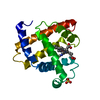
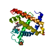
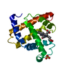
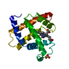
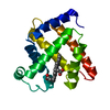
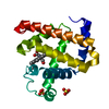
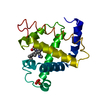
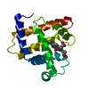
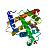
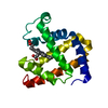
| Entry | Database: PDB / ID: 4mbn | ||||||||||||
|---|---|---|---|---|---|---|---|---|---|---|---|---|---|
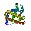
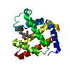
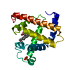
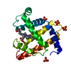
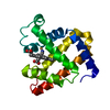
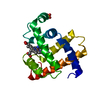
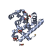
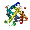
| Title | REFINEMENT OF MYOGLOBIN AND CYTOCHROME C | ||||||||||||
 Components Components | MYOGLOBIN | ||||||||||||
 Keywords Keywords | OXYGEN STORAGE | ||||||||||||
| Function / homology |  Function and homology information Function and homology informationOxidoreductases; Acting on other nitrogenous compounds as donors / nitrite reductase activity / sarcoplasm / Oxidoreductases; Acting on a peroxide as acceptor; Peroxidases / removal of superoxide radicals / oxygen carrier activity / peroxidase activity / oxygen binding / heme binding / extracellular exosome / metal ion binding Similarity search - Function | ||||||||||||
| Biological species |  | ||||||||||||
| Method |  X-RAY DIFFRACTION / Resolution: 2 Å X-RAY DIFFRACTION / Resolution: 2 Å | ||||||||||||
 Authors Authors | Takano, T. | ||||||||||||
 Citation Citation | Journal: Methods and Applications in Crystallographic Computing Year: 1984 Title: Refinement of Myoglobin and Cytochrome C Authors: Takano, T. #1: Journal: J.Mol.Biol. / Year: 1977Title: Structure of Myoglobin Refined at 2.0 Angstroms Resolution. II. Structure of Deoxymyoglobin from Sperm Whale Authors: Takano, T. #2: Journal: J.Mol.Biol. / Year: 1977Title: Structure of Myoglobin Refined at 2.0 Angstroms Resolution. I. Crystallographic Refinement of Metmyoglobin from Sperm Whale Authors: Takano, T. #3: Journal: Prog.Stereochem. / Year: 1976Title: The Stereochemistry of the Protein Myoglobin Authors: Watson, H.C. #4: Journal: Acta Crystallogr. / Year: 1966Title: A Mathematical Model-Building Procedure for Proteins Authors: Diamond, R. #5: Journal: Acta Crystallogr.,Sect.A / Year: 1971Title: A Real-Space Refinement Procedure for Proteins Authors: Diamond, R. #6: Journal: J.Mol.Biol. / Year: 1974Title: Real-Space Refinement of the Structure of Hen Egg-White Lysozyme Authors: Diamond, R. #7: Journal: Nature / Year: 1966Title: Structure of Deoxymyoglobin, a Crystallographic Study Authors: Nobbs, C.L. / Watson, H.C. / Kendrew, J.C. #8: Journal: J.Mol.Biol. / Year: 1975Title: Three-Dimensional Fourier Synthesis of Human Deoxyhaemoglobin at 2.5 Angstroms, Refinement of the Atomic Model Authors: Fermi, G. | ||||||||||||
| History |
|
- Structure visualization
| Structure viewer | Molecule: MolmilJmol/JSmol |
|---|
- Downloads & links
-Download
| PDBx/mmCIF format | 4mbn.cif.gz | 46.4 KB | Display | PDBx/mmCIF format |
|---|---|---|---|---|
| PDB format | pdb4mbn.ent.gz | 32.8 KB | Display | PDB format |
| PDBx/mmJSON format | 4mbn.json.gz | Tree view | PDBx/mmJSON format | |
| Others |  Other downloads Other downloads |
-Validation report
| Arichive directory | https://data.pdbj.org/pub/pdb/validation_reports/mb/4mbnftp://data.pdbj.org/pub/pdb/validation_reports/mb/4mbn | HTTPS FTP |
|---|
-Related structure data
| Similar structure data |
|---|


-Links
 PDBj
PDBj


- Assembly
| Deposited unit | 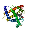
| ||||||||
|---|---|---|---|---|---|---|---|---|---|
| 1 |
| ||||||||
| Unit cell |
| ||||||||
| Atom site foot note | 1: SEE REMARK 5. |
-Components
| #1: Protein | Mass: 17234.951 Da / Num. of mol.: 1 Source method: isolated from a genetically manipulated source Source: (gene. exp.) | ||||
|---|---|---|---|---|---|
| #2: Chemical |   Mass: 96.063 Da / Num. of mol.: 2 / Source method: obtained synthetically / Formula: SO4 Mass: 96.063 Da / Num. of mol.: 2 / Source method: obtained synthetically / Formula: SO4#3: Chemical | ChemComp-HEM / |   Mass: 616.487 Da / Num. of mol.: 1 / Source method: obtained synthetically / Formula: C34H32FeN4O4 Mass: 616.487 Da / Num. of mol.: 1 / Source method: obtained synthetically / Formula: C34H32FeN4O4#4: Water | ChemComp-HOH / |  Mass: 18.015 Da / Num. of mol.: 121 / Source method: isolated from a natural source / Formula: H2O Mass: 18.015 Da / Num. of mol.: 121 / Source method: isolated from a natural source / Formula: H2O |
-Experimental details
-Experiment
| Experiment | Method: X-RAY DIFFRACTION |
|---|
- Sample preparation
| Crystal | Density Matthews: 1.94 Å3/Da / Density % sol: 36.75 % |
|---|
- Processing
| Software | Name: EREF / Classification: refinement | ||||||||||||
|---|---|---|---|---|---|---|---|---|---|---|---|---|---|
| Refinement | Rfactor Rwork: 0.172 / Highest resolution: 2 Å | ||||||||||||
| Refinement step | Cycle: LAST / Highest resolution: 2 Å
|
