 Movie
Movie Controller

[English] 日本語
 Yorodumi
- PDB-1l2k: Neutron Structure Determination of Sperm Whale Met-Myoglobin at 1... -
+ Open data

- Basic information
| Entry | Database: PDB / ID: 1l2k | ||||||
|---|---|---|---|---|---|---|---|
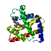
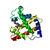
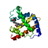
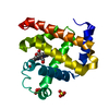
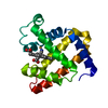
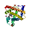
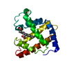
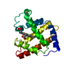
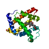
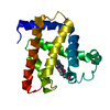
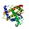
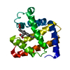
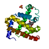
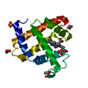
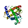
| Title | Neutron Structure Determination of Sperm Whale Met-Myoglobin at 1.5A Resolution. | ||||||
 Components Components | MYOGLOBIN | ||||||
 Keywords Keywords | OXYGEN STORAGE/TRANSPORT / NEUTRON STRUCTURE / HYDROGEN ATOMS / HYDRATION STRUCTURE / HEME PROTEIN / OXYGEN STORAGE-TRANSPORT COMPLEX | ||||||
| Function / homology |  Function and homology information Function and homology informationOxidoreductases; Acting on other nitrogenous compounds as donors / nitrite reductase activity / sarcoplasm / Oxidoreductases; Acting on a peroxide as acceptor; Peroxidases / removal of superoxide radicals / oxygen carrier activity / peroxidase activity / oxygen binding / heme binding / extracellular exosome / metal ion binding Similarity search - Function | ||||||
| Biological species |  | ||||||
| Method | NEUTRON DIFFRACTION / NUCLEAR REACTOR / Resolution: 1.5 Å | ||||||
 Authors Authors | Ostermann, A. / Tanaka, I. / Engler, N. / Niimura, N. / Parak, F.G. | ||||||
 Citation Citation | Journal: Biophys.Chem. / Year: 2002 Title: Hydrogen and deuterium in myoglobin as seen by a neutron structure determination at 1.5 A resolution. Authors: Ostermann, A. / Tanaka, I. / Engler, N. / Niimura, N. / Parak, F.G. #1: Journal: CURR.OPIN.STRUCT.BIOL. / Year: 1999Title: Neutrons Expand the Field of Structural Biology Authors: Niimura, N. | ||||||
| History |
|
- Structure visualization
| Structure viewer | Molecule: MolmilJmol/JSmol |
|---|
- Downloads & links
-Download
| PDBx/mmCIF format | 1l2k.cif.gz | 74.9 KB | Display | PDBx/mmCIF format |
|---|---|---|---|---|
| PDB format | pdb1l2k.ent.gz | 57.9 KB | Display | PDB format |
| PDBx/mmJSON format | 1l2k.json.gz | Tree view | PDBx/mmJSON format | |
| Others |  Other downloads Other downloads |
-Validation report
| Arichive directory | https://data.pdbj.org/pub/pdb/validation_reports/l2/1l2kftp://data.pdbj.org/pub/pdb/validation_reports/l2/1l2k | HTTPS FTP |
|---|
-Related structure data
| Similar structure data |
|---|


-Links
 PDBj
PDBj


- Assembly
| Deposited unit | 
| ||||||||
|---|---|---|---|---|---|---|---|---|---|
| 1 |
| ||||||||
| Unit cell |
|
-Components
| #1: Protein | Mass: 17234.951 Da / Num. of mol.: 1 / Source method: isolated from a natural source / Source: (natural) | ||||||
|---|---|---|---|---|---|---|---|
| #2: Chemical |   Mass: 96.063 Da / Num. of mol.: 2 / Source method: obtained synthetically / Formula: SO4 Mass: 96.063 Da / Num. of mol.: 2 / Source method: obtained synthetically / Formula: SO4#3: Chemical | ChemComp-ND4 / |   Mass: 22.063 Da / Num. of mol.: 1 / Source method: obtained synthetically / Formula: N Mass: 22.063 Da / Num. of mol.: 1 / Source method: obtained synthetically / Formula: N#4: Chemical | ChemComp-HEM / |   Mass: 616.487 Da / Num. of mol.: 1 / Source method: obtained synthetically / Formula: C34H32FeN4O4 Mass: 616.487 Da / Num. of mol.: 1 / Source method: obtained synthetically / Formula: C34H32FeN4O4#5: Chemical | ChemComp-DOD / |   Mass: 18.015 Da / Num. of mol.: 74 / Source method: isolated from a natural source / Formula: D2O Mass: 18.015 Da / Num. of mol.: 74 / Source method: isolated from a natural source / Formula: D2O |
-Experimental details
-Experiment
| Experiment | Method: NEUTRON DIFFRACTION / Number of used crystals: 1 |
|---|
- Sample preparation
| Crystal grow | Temperature: 298 K / Method: batch crystallization / pH: 6.8 Details: AMMONIUM SULFATE, POTASSIUM PHOSPHATE, pH 6.8, BATCH CRYSTALLIZATION, temperature 298K | |||||||||||||||||||||
|---|---|---|---|---|---|---|---|---|---|---|---|---|---|---|---|---|---|---|---|---|---|---|
| Crystal grow | *PLUS Method: unknown | |||||||||||||||||||||
| Components of the solutions | *PLUS
|
-Data collection
| Diffraction | Mean temperature: 298 K |
|---|---|
| Diffraction source | Source: NUCLEAR REACTOR / Beamline: BIX-3 (1G-A BEAM PORT) / Type: JRR-3M, GUIDE 1G-A, BIX-3 / Wavelength: 2.35 Å |
| Detector | Type: MACSCIENCE / Detector: NEUTRON IMAGE PLATE / Date: Feb 5, 2000 |
| Radiation | Monochromator: ELASTICALLY BENT SILICON / Protocol: SINGLE WAVELENGTH / Monochromatic (M) / Laue (L): M / Scattering type: x-ray |
| Radiation wavelength | Wavelength: 2.35 Å / Relative weight: 1 |
| Reflection | Resolution: 1.5→25 Å / Num. obs: 19135 / % possible obs: 87.9 % / Redundancy: 2.9 % / Biso Wilson estimate: 8.5 Å2 / Rmerge(I) obs: 0.103 / Net I/σ(I): 6.3 |
| Reflection shell | Resolution: 1.5→1.55 Å / Redundancy: 2.1 % / Rmerge(I) obs: 0.249 / Mean I/σ(I) obs: 2.7 / Num. unique all: 1458 / % possible all: 67 |
| Reflection | *PLUS Lowest resolution: 22 Å / Num. measured all: 55899 / Rmerge(I) obs: 0.103 |
| Reflection shell | *PLUS % possible obs: 67 % / Num. unique obs: 1458 / Num. measured obs: 3041 / Rmerge(I) obs: 0.249 |
- Processing
| Software |
| ||||||||||||||||||||||||||||||||||||||||||||||||||||||||||||||||||||||||||||||||
|---|---|---|---|---|---|---|---|---|---|---|---|---|---|---|---|---|---|---|---|---|---|---|---|---|---|---|---|---|---|---|---|---|---|---|---|---|---|---|---|---|---|---|---|---|---|---|---|---|---|---|---|---|---|---|---|---|---|---|---|---|---|---|---|---|---|---|---|---|---|---|---|---|---|---|---|---|---|---|---|---|---|
| Refinement | Starting model: X-RAY STRUCTURE Resolution: 1.5→22.7 Å / Rfactor Rfree error: 0.006 / Data cutoff high absF: 10000000 / Data cutoff low absF: 0 / Isotropic thermal model: RESTRAINED / Cross valid method: THROUGHOUT / σ(F): 0 Stereochemistry target values: MAXIMUM LIKELIHOOD TARGET USING AMPLITUDES Details: X-PLOR 3.851 was also used in refinement. THE STANDARD TOPOLOGY AND PARAMETER FILES WERE CHANGED FOR SEVERAL HYDROGEN ATOM PARAMETERS TO MEET THE REQUIREMENTS OF THE NEUTRON STRUCTURE ...Details: X-PLOR 3.851 was also used in refinement. THE STANDARD TOPOLOGY AND PARAMETER FILES WERE CHANGED FOR SEVERAL HYDROGEN ATOM PARAMETERS TO MEET THE REQUIREMENTS OF THE NEUTRON STRUCTURE REFINEMENT. THE FOLLOWING NEUTRON-SCATTERING LENGTHS WERE USED FOR THE REFINEMENT: ATOM H = -0.374 10**-12 CM. ATOM D = 0.667 10**-12 CM. ATOM C = 0.665 10**-12 CM. ATOM N = 0.921 10**-12 CM. ATOM O = 0.581 10**-12 CM. ATOM S = 0.285 10**-12 CM. ATOM FE = 0.954 10**-12 CM. DEUTERIUM ATOMS IN AMINO ACID SIDE CHAINS WERE ONLY INCLUDED INTO THE MODEL IF A SIGNIFICANT DENSITY FEATURE WAS PRESENT. OCCUPANCIES FOR THE BACKBONE AMIDE HYDROGEN ATOMS WERE REFINED (H/D EXCHANGE). FOR THE OCCUPANCY REFINEMENT NO CONSTRAINT FOR ADDING UP THE OCCUPANCIES TO 1.0 WAS USED. THE ADDED FRACTIONAL OCCUPANCY AVERAGED OVER ALL BACKBONE AMIDE GROUPS YIELDS A VALUE OF 1.09 WITH AN S.D. OF 0.125. THE VALUES GIVEN IN THIS COORDINATE FILE ARE NORMALIZED. A POSITIONAL REFINEMENT FOR THE BACKBONE AMIDE HYDROGEN ATOMS WITH WEAKENED IN-PLANE RESTRAINTS FOR THE HYDROGEN ATOM WITH RESPECT TO THE AMIDE PLANE SHOWED DEVIATIONS GREATER THAN 10 DEGREE FOR THE FOLLOWING RESIDUES: 12,15, 31,48,51,56,58,80,94,96,97,99,101,103,104,107,144. THE COORDINATES GIVEN IN THIS FILE WERE REFINED WITH NORMAL RESTRAINTS. IN HIS 97 THE HYDROGEN ATOM HE1 WHICH IS BOUND TO THE CARBON ATOM CE1 IS EXCHANGED TO DEUTERIUM. FOR SEVERAL WATER MOLECULES (DOD) ONLY THE O-ATOM AND ONE D-ATOM COULD BE OBSERVED IN THE DENSITY MAP. THE SECOND D-ATOM IS STRONGLY DISORDERED. THESE WATER MOLECULES WERE MODELED AS OD. IT DOES NOT MEAN HYDROXYL-ION. THERE IS NEARLY NO NEUTRON DENSITY FOR RESIDUE 152 AND 153. THOSE RESIDUES WERE NOT INCLUDED INTO THE MODEL.
| ||||||||||||||||||||||||||||||||||||||||||||||||||||||||||||||||||||||||||||||||
| Solvent computation | Solvent model: flat model / Bsol: 120 Å2 / ksol: 0.0625 e/Å3 | ||||||||||||||||||||||||||||||||||||||||||||||||||||||||||||||||||||||||||||||||
| Displacement parameters | Biso mean: 12.9 Å2 | ||||||||||||||||||||||||||||||||||||||||||||||||||||||||||||||||||||||||||||||||
| Refine analyze |
| ||||||||||||||||||||||||||||||||||||||||||||||||||||||||||||||||||||||||||||||||
| Refinement step | Cycle: LAST / Resolution: 1.5→22.7 Å
| ||||||||||||||||||||||||||||||||||||||||||||||||||||||||||||||||||||||||||||||||
| Refine LS restraints |
| ||||||||||||||||||||||||||||||||||||||||||||||||||||||||||||||||||||||||||||||||
| LS refinement shell | Resolution: 1.5→1.55 Å / Rfactor Rfree error: 0.03 / Total num. of bins used: 10
| ||||||||||||||||||||||||||||||||||||||||||||||||||||||||||||||||||||||||||||||||
| Xplor file |
| ||||||||||||||||||||||||||||||||||||||||||||||||||||||||||||||||||||||||||||||||
| Refinement | *PLUS Highest resolution: 1.5 Å / Lowest resolution: 22 Å / Num. reflection obs: 17824 / Num. reflection Rfree: 1311 / Rfactor Rfree: 0.247 / Rfactor Rwork: 0.214 | ||||||||||||||||||||||||||||||||||||||||||||||||||||||||||||||||||||||||||||||||
| Solvent computation | *PLUS | ||||||||||||||||||||||||||||||||||||||||||||||||||||||||||||||||||||||||||||||||
| Displacement parameters | *PLUS | ||||||||||||||||||||||||||||||||||||||||||||||||||||||||||||||||||||||||||||||||
| Refine LS restraints | *PLUS
| ||||||||||||||||||||||||||||||||||||||||||||||||||||||||||||||||||||||||||||||||
| LS refinement shell | *PLUS Rfactor Rfree: 0.3057 / Rfactor Rwork: 0.2825 |
 X-PLOR