 Movie
Movie Controller

[English] 日本語
 Yorodumi
- PDB-2mb5: HYDRATION IN PROTEIN CRYSTALS. A NEUTRON DIFFRACTION ANALYSIS OF ... -
+ Open data

- Basic information
| Entry | Database: PDB / ID: 2mb5 | |||||||||
|---|---|---|---|---|---|---|---|---|---|---|
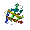
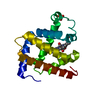
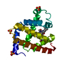
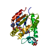
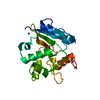
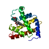
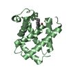
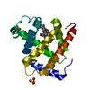
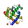
| Title | HYDRATION IN PROTEIN CRYSTALS. A NEUTRON DIFFRACTION ANALYSIS OF CARBONMONOXYMYOGLOBIN | |||||||||
 Components Components | MYOGLOBIN | |||||||||
 Keywords Keywords | OXYGEN STORAGE | |||||||||
| Function / homology |  Function and homology information Function and homology informationOxidoreductases; Acting on other nitrogenous compounds as donors / nitrite reductase activity / sarcoplasm / Oxidoreductases; Acting on a peroxide as acceptor; Peroxidases / removal of superoxide radicals / oxygen carrier activity / peroxidase activity / oxygen binding / heme binding / extracellular exosome / metal ion binding Similarity search - Function | |||||||||
| Biological species |  | |||||||||
| Method | NEUTRON DIFFRACTION / Resolution: 1.8 Å | |||||||||
 Authors Authors | Schoenborn, B.P. / Cheng, X. | |||||||||
 Citation Citation | Journal: Acta Crystallogr.,Sect.B / Year: 1990 Title: Hydration in Protein Crystals. A Neutron Diffraction Analysis of Carbonmonoxymyoglobin Authors: Cheng, X. / Schoenborn, B.P. #1: Journal: J.Mol.Biol. / Year: 1981Title: Real Space Refinement of Neutron Diffraction Data from Sperm Whale Carbonmonoxymyoglobin Authors: Hanson, J.C. / Schoenborn, B.P. #2: Journal: Acs Symp.Ser. / Year: 1980Title: The Determination of Structural Water by Neutron Protein Crystallography. An Analysis of the Carbon Monoxide Myoglobin Water Structure Authors: Schoenborn, B.P. / Hanson, J.C. #3: Journal: Acta Crystallogr.,Sect.A (Supplement) / Year: 1975Title: Neutron Diffraction Analysis and Real Space Refinement of met and Carbon Monoxide Myoglobin Authors: Schoenborn, B.P. / Norvell, J.C. #4: Journal: Brookhaven Symposia in Biology / Year: 1975Title: Neutron Diffraction Analysis of Metmyoglobin Authors: Schoenborn, B.P. / Diamond, R. #5: Journal: Science / Year: 1975Title: Neutron Diffraction Analysis of Myoglobin. Structure of the Carbon Monoxide Derivative Authors: Norvell, J.C. / Nunes, A.C. / Schoenborn, B.P. #6: Journal: Cold Spring Harbor Symp.Quant.Biol. / Year: 1972Title: A Neutron Diffraction Analysis of Myoglobin. III. Hydrogen-Deuterium Bonding in Side Chains Authors: Schoenborn, B.P. #7: Journal: Nature / Year: 1969Title: Neutron Diffraction Analysis of Myoglobin Authors: Schoenborn, B.P. | |||||||||
| History |
|
- Structure visualization
| Structure viewer | Molecule: MolmilJmol/JSmol |
|---|
- Downloads & links
-Download
| PDBx/mmCIF format | 2mb5.cif.gz | 79.2 KB | Display | PDBx/mmCIF format |
|---|---|---|---|---|
| PDB format | pdb2mb5.ent.gz | 62.4 KB | Display | PDB format |
| PDBx/mmJSON format | 2mb5.json.gz | Tree view | PDBx/mmJSON format | |
| Others |  Other downloads Other downloads |
-Validation report
| Arichive directory | https://data.pdbj.org/pub/pdb/validation_reports/mb/2mb5ftp://data.pdbj.org/pub/pdb/validation_reports/mb/2mb5 | HTTPS FTP |
|---|
-Related structure data
| Similar structure data |
|---|

-Links
 PDBj
PDBj


- Assembly
| Deposited unit | 
| ||||||||
|---|---|---|---|---|---|---|---|---|---|
| 1 |
| ||||||||
| Unit cell |
|
-Components
-Protein , 1 types, 1 molecules A
| #1: Protein | Mass: 17234.951 Da / Num. of mol.: 1 Source method: isolated from a genetically manipulated source Source: (gene. exp.) |
|---|
-Non-polymers , 5 types, 97 molecules 


| #2: Chemical | ChemComp-SO4 /  Mass: 96.063 Da / Num. of mol.: 1 / Source method: obtained synthetically / Formula: SO4 Mass: 96.063 Da / Num. of mol.: 1 / Source method: obtained synthetically / Formula: SO4 | ||||||
|---|---|---|---|---|---|---|---|
| #3: Chemical | ChemComp-ND4 /  Mass: 22.063 Da / Num. of mol.: 5 / Source method: obtained synthetically / Formula: N Mass: 22.063 Da / Num. of mol.: 5 / Source method: obtained synthetically / Formula: N#4: Chemical | ChemComp-HEM / |  Mass: 616.487 Da / Num. of mol.: 1 / Source method: obtained synthetically / Formula: C34H32FeN4O4 Mass: 616.487 Da / Num. of mol.: 1 / Source method: obtained synthetically / Formula: C34H32FeN4O4#5: Chemical | ChemComp-CMO / |  Mass: 28.010 Da / Num. of mol.: 1 / Source method: obtained synthetically / Formula: CO Mass: 28.010 Da / Num. of mol.: 1 / Source method: obtained synthetically / Formula: CO#6: Chemical | ChemComp-DOD / |  Mass: 18.015 Da / Num. of mol.: 89 / Source method: isolated from a natural source / Formula: D2O Mass: 18.015 Da / Num. of mol.: 89 / Source method: isolated from a natural source / Formula: D2O |
-Experimental details
-Experiment
| Experiment | Method: NEUTRON DIFFRACTION |
|---|
- Sample preparation
| Crystal grow | *PLUS Method: unknown |
|---|
- Processing
| Software | Name: PROLSQ / Classification: refinement | ||||||||||||
|---|---|---|---|---|---|---|---|---|---|---|---|---|---|
| Refinement | Highest resolution: 1.8 Å | ||||||||||||
| Refinement step | Cycle: LAST / Highest resolution: 1.8 Å
| ||||||||||||
| Refine LS restraints |
|
