 Movie
Movie Controller

[English] 日本語
 Yorodumi
- PDB-1qhy: CHLORAMPHENICOL PHOSPHOTRANSFERASE FROM STREPTOMYCES VENEZUELAE I... -
+ Open data

- Basic information
| Entry | Database: PDB / ID: 1qhy | ||||||
|---|---|---|---|---|---|---|---|
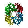
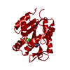
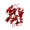
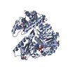
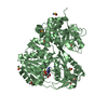
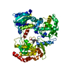
| Title | CHLORAMPHENICOL PHOSPHOTRANSFERASE FROM STREPTOMYCES VENEZUELAE IN COMPLEX WITH ATPGAMMAS AND CHLORAMPHENICOL | ||||||
 Components Components | CHLORAMPHENICOL PHOSPHOTRANSFERASE | ||||||
 Keywords Keywords | TRANSFERASE / KINASE / ANTIBIOTIC RESISTANCE / PHOSPHORYLATION / MONONUCLEOTIDE BINDING FOLD | ||||||
| Function / homology |  Function and homology information Function and homology informationTransferases; Transferring phosphorus-containing groups; Phosphotransferases with an alcohol group as acceptor / kinase activity / response to antibiotic / ATP binding Similarity search - Function | ||||||
| Biological species |  Streptomyces venezuelae (bacteria) Streptomyces venezuelae (bacteria) | ||||||
| Method |  X-RAY DIFFRACTION / MOLECULAR REPLACEMENT / Resolution: 2.6 Å X-RAY DIFFRACTION / MOLECULAR REPLACEMENT / Resolution: 2.6 Å | ||||||
 Authors Authors | Izard, T. | ||||||
 Citation Citation | Journal: Embo J. / Year: 2000 Title: The Crystal Structures of Chloramphenicol Phosphotransferase Reveal a Novel Inactivation Mechanism Authors: Izard, T. / Ellis, J. | ||||||
| History |
|
- Structure visualization
| Structure viewer | Molecule: MolmilJmol/JSmol |
|---|
- Downloads & links
-Download
| PDBx/mmCIF format | 1qhy.cif.gz | 41.5 KB | Display | PDBx/mmCIF format |
|---|---|---|---|---|
| PDB format | pdb1qhy.ent.gz | 33.3 KB | Display | PDB format |
| PDBx/mmJSON format | 1qhy.json.gz | Tree view | PDBx/mmJSON format | |
| Others |  Other downloads Other downloads |
-Validation report
| Arichive directory | https://data.pdbj.org/pub/pdb/validation_reports/qh/1qhyftp://data.pdbj.org/pub/pdb/validation_reports/qh/1qhy | HTTPS FTP |
|---|
-Related structure data
| Related structure data | 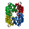 1qhnSC 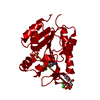 1qhsC  1qhxC S: Starting model for refinement C: citing same article ( |
|---|---|
| Similar structure data |


-Links
 PDBj
PDBj
- Assembly
| Deposited unit | 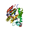
| ||||||||
|---|---|---|---|---|---|---|---|---|---|
| 1 | 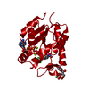
| ||||||||
| Unit cell |
| ||||||||
| Components on special symmetry positions |
|
-Components
-Protein , 1 types, 1 molecules A
| #1: Protein | Mass: 18834.365 Da / Num. of mol.: 1 / Source method: isolated from a natural source / Source: (natural) Streptomyces venezuelae (bacteria) / Strain: ISP5230References: UniProt: Q56148, Transferases; Transferring phosphorus-containing groups; Phosphotransferases with an alcohol group as acceptor |
|---|
-Non-polymers , 5 types, 50 molecules 


| #2: Chemical | ChemComp-SO4 /  Mass: 96.063 Da / Num. of mol.: 1 / Source method: obtained synthetically / Formula: SO4 Mass: 96.063 Da / Num. of mol.: 1 / Source method: obtained synthetically / Formula: SO4 | ||
|---|---|---|---|
| #3: Chemical | ChemComp-MG /  Mass: 24.305 Da / Num. of mol.: 1 / Source method: obtained synthetically / Formula: Mg Mass: 24.305 Da / Num. of mol.: 1 / Source method: obtained synthetically / Formula: Mg | ||
| #4: Chemical | ChemComp-AGS /  Mass: 523.247 Da / Num. of mol.: 1 / Source method: obtained synthetically / Formula: C10H16N5O12P3S / Comment: ATP-gamma-S, energy-carrying molecule analogue*YM Mass: 523.247 Da / Num. of mol.: 1 / Source method: obtained synthetically / Formula: C10H16N5O12P3S / Comment: ATP-gamma-S, energy-carrying molecule analogue*YM | ||
| #5: Chemical |  Mass: 323.129 Da / Num. of mol.: 2 / Source method: obtained synthetically / Formula: C11H12Cl2N2O5 / Comment: antibiotic*YM Mass: 323.129 Da / Num. of mol.: 2 / Source method: obtained synthetically / Formula: C11H12Cl2N2O5 / Comment: antibiotic*YM#6: Water | ChemComp-HOH / | Mass: 18.015 Da / Num. of mol.: 45 / Source method: isolated from a natural source / Formula: H2O |
-Experimental details
-Experiment
| Experiment | Method: X-RAY DIFFRACTION / Number of used crystals: 1 |
|---|
- Sample preparation
| Crystal | Density Matthews: 8.8 Å3/Da / Density % sol: 86 % | ||||||||||||||||||||||||||||||||||||||||
|---|---|---|---|---|---|---|---|---|---|---|---|---|---|---|---|---|---|---|---|---|---|---|---|---|---|---|---|---|---|---|---|---|---|---|---|---|---|---|---|---|---|
| Crystal grow | pH: 7.5 / Details: pH 7.50 | ||||||||||||||||||||||||||||||||||||||||
| Crystal grow | *PLUS Method: vapor diffusion | ||||||||||||||||||||||||||||||||||||||||
| Components of the solutions | *PLUS
|
-Data collection
| Diffraction | Mean temperature: 100 K |
|---|---|
| Diffraction source | Source: ROTATING ANODE / Wavelength: 1.54 |
| Detector | Detector: IMAGE PLATE |
| Radiation | Protocol: SINGLE WAVELENGTH / Monochromatic (M) / Laue (L): M / Scattering type: x-ray |
| Radiation wavelength | Wavelength: 1.54 Å / Relative weight: 1 |
| Reflection | Resolution: 2.6→20 Å / Num. obs: 21331 / % possible obs: 98.1 % / Redundancy: 14.52 % / Rmerge(I) obs: 0.061 / Rsym value: 0.064 / Net I/σ(I): 16.2 |
| Reflection | *PLUS % possible obs: 99.7 % / Redundancy: 14.26 % / Num. measured all: 304201 |
| Reflection shell | *PLUS % possible obs: 99.8 % / Rmerge(I) obs: 0.367 |
- Processing
| Software |
| ||||||||||||||||||||||||||||||||||||||||||||||||||||||||||||
|---|---|---|---|---|---|---|---|---|---|---|---|---|---|---|---|---|---|---|---|---|---|---|---|---|---|---|---|---|---|---|---|---|---|---|---|---|---|---|---|---|---|---|---|---|---|---|---|---|---|---|---|---|---|---|---|---|---|---|---|---|---|
| Refinement | Method to determine structure: MOLECULAR REPLACEMENT Starting model: 1QHN Resolution: 2.6→20 Å / Cross valid method: THROUGHOUT / σ(F): 0
| ||||||||||||||||||||||||||||||||||||||||||||||||||||||||||||
| Refinement step | Cycle: LAST / Resolution: 2.6→20 Å
| ||||||||||||||||||||||||||||||||||||||||||||||||||||||||||||
| Refine LS restraints |
| ||||||||||||||||||||||||||||||||||||||||||||||||||||||||||||
| Software | *PLUS Name: CNS / Version: 0.5 / Classification: refinement | ||||||||||||||||||||||||||||||||||||||||||||||||||||||||||||
| LS refinement shell | *PLUS Rfactor Rfree: 0.358 / Rfactor Rwork: 0.349 |
