 Movie
Movie Controller

[English] 日本語
 Yorodumi
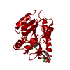
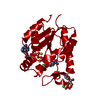
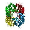
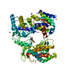
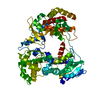
- PDB-1grq: CHLORAMPHENICOL PHOSPHOTRANSFERASE IN COMPLEX WITH P-AMINO-CHLORA... -
+ Open data

- Basic information
| Entry | Database: PDB / ID: 1grq | ||||||
|---|---|---|---|---|---|---|---|
| Title | CHLORAMPHENICOL PHOSPHOTRANSFERASE IN COMPLEX WITH P-AMINO-CHLORAMPHENICOL FROM STREPTOMYCES VENEZUELAE | ||||||
 Components Components | CHLORAMPHENICOL 3-O PHOSPHOTRANSFERASE | ||||||
 Keywords Keywords | TRANSFERASE / KINASE / ANTIBIOTIC RESISTANCE / PHOSPHORYLATION / MONONUCLEOTIDE BINDING FOLD | ||||||
| Function / homology |  Function and homology information Function and homology informationTransferases; Transferring phosphorus-containing groups; Phosphotransferases with an alcohol group as acceptor / kinase activity / response to antibiotic / ATP binding Similarity search - Function | ||||||
| Biological species |  STREPTOMYCES VENEZUELAE (bacteria) STREPTOMYCES VENEZUELAE (bacteria) | ||||||
| Method |  X-RAY DIFFRACTION / SYNCHROTRON / MOLECULAR REPLACEMENT / Resolution: 2.9 Å X-RAY DIFFRACTION / SYNCHROTRON / MOLECULAR REPLACEMENT / Resolution: 2.9 Å | ||||||
 Authors Authors | Izard, T. | ||||||
 Citation Citation | Journal: Protein Sci. / Year: 2001 Title: Structural Basis for Chloramphenicol Tolerance in Streptomyces Venezuelae by Chloramphenicol Phosphotransferase Activity Authors: Izard, T. | ||||||
| History |
|
- Structure visualization
| Structure viewer | Molecule: MolmilJmol/JSmol |
|---|
- Downloads & links
-Download
| PDBx/mmCIF format | 1grq.cif.gz | 44.8 KB | Display | PDBx/mmCIF format |
|---|---|---|---|---|
| PDB format | pdb1grq.ent.gz | 32.7 KB | Display | PDB format |
| PDBx/mmJSON format | 1grq.json.gz | Tree view | PDBx/mmJSON format | |
| Others |  Other downloads Other downloads |
-Validation report
| Arichive directory | https://data.pdbj.org/pub/pdb/validation_reports/gr/1grqftp://data.pdbj.org/pub/pdb/validation_reports/gr/1grq | HTTPS FTP |
|---|
-Related structure data
| Related structure data | 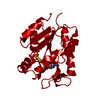 1grrC 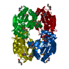 1qhnS C: citing same article ( S: Starting model for refinement |
|---|---|
| Similar structure data |


-Links
 PDBj
PDBj- Assembly
| Deposited unit | 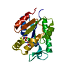
| ||||||||
|---|---|---|---|---|---|---|---|---|---|
| 1 | 
| ||||||||
| Unit cell |
|
-Components
| #1: Protein | Mass: 18834.365 Da / Num. of mol.: 1 / Source method: isolated from a natural source / Source: (natural) STREPTOMYCES VENEZUELAE (bacteria) / Strain: ISP5230 / References: UniProt: Q56148 |
|---|---|
| #2: Chemical | ChemComp-SO4 /   Mass: 96.063 Da / Num. of mol.: 1 / Source method: obtained synthetically / Formula: SO4 Mass: 96.063 Da / Num. of mol.: 1 / Source method: obtained synthetically / Formula: SO4 |
| #3: Chemical | ChemComp-CLK /   Mass: 293.146 Da / Num. of mol.: 1 / Source method: obtained synthetically / Formula: C11H14Cl2N2O3 Mass: 293.146 Da / Num. of mol.: 1 / Source method: obtained synthetically / Formula: C11H14Cl2N2O3 |
| #4: Water | ChemComp-HOH /  Mass: 18.015 Da / Num. of mol.: 92 / Source method: isolated from a natural source / Formula: H2O Mass: 18.015 Da / Num. of mol.: 92 / Source method: isolated from a natural source / Formula: H2O |
| Has protein modification | N |
-Experimental details
-Experiment
| Experiment | Method: X-RAY DIFFRACTION / Number of used crystals: 1 |
|---|
- Sample preparation
| Crystal | Density Matthews: 8.8 Å3/Da / Density % sol: 86 % | ||||||||||||||||||||||||||||||||||||||||||||||||
|---|---|---|---|---|---|---|---|---|---|---|---|---|---|---|---|---|---|---|---|---|---|---|---|---|---|---|---|---|---|---|---|---|---|---|---|---|---|---|---|---|---|---|---|---|---|---|---|---|---|
| Crystal grow | pH: 7.5 / Details: pH 7.50 | ||||||||||||||||||||||||||||||||||||||||||||||||
| Crystal grow | *PLUS pH: 7.5 / Method: vapor diffusion, hanging drop | ||||||||||||||||||||||||||||||||||||||||||||||||
| Components of the solutions | *PLUS
|
-Data collection
| Diffraction | Mean temperature: 100 K |
|---|---|
| Diffraction source | Source: SYNCHROTRON / Site: SRS  / Beamline: PX9.6 / Wavelength: 0.87 / Beamline: PX9.6 / Wavelength: 0.87 |
| Detector | Detector: CCD |
| Radiation | Protocol: SINGLE WAVELENGTH / Monochromatic (M) / Laue (L): M / Scattering type: x-ray |
| Radiation wavelength | Wavelength: 0.87 Å / Relative weight: 1 |
| Reflection | Resolution: 2.9→100 Å / Num. obs: 370300 / % possible obs: 95.1 % / Observed criterion σ(I): 2 / Redundancy: 25.1 % / Rmerge(I) obs: 0.051 / Net I/σ(I): 48.8 |
| Reflection shell | Resolution: 2.9→3 Å / Rmerge(I) obs: 0.298 / % possible all: 0.6 |
| Reflection | *PLUS Num. obs: 14719 / Num. measured all: 370300 |
| Reflection shell | *PLUS % possible obs: 59.3 % |
- Processing
| Software |
| ||||||||||||||||||||||||||||||||||||||||||||||||||||||||||||
|---|---|---|---|---|---|---|---|---|---|---|---|---|---|---|---|---|---|---|---|---|---|---|---|---|---|---|---|---|---|---|---|---|---|---|---|---|---|---|---|---|---|---|---|---|---|---|---|---|---|---|---|---|---|---|---|---|---|---|---|---|---|
| Refinement | Method to determine structure: MOLECULAR REPLACEMENT Starting model: 1QHN Resolution: 2.9→99 Å / Rfactor Rfree error: 0.011 / Cross valid method: THROUGHOUT / σ(F): 0
| ||||||||||||||||||||||||||||||||||||||||||||||||||||||||||||
| Displacement parameters |
| ||||||||||||||||||||||||||||||||||||||||||||||||||||||||||||
| Refine analyze | Luzzati coordinate error free: 0.43 Å / Luzzati sigma a free: 0.49 Å | ||||||||||||||||||||||||||||||||||||||||||||||||||||||||||||
| Refinement step | Cycle: LAST / Resolution: 2.9→99 Å
| ||||||||||||||||||||||||||||||||||||||||||||||||||||||||||||
| Refine LS restraints |
| ||||||||||||||||||||||||||||||||||||||||||||||||||||||||||||
| LS refinement shell | Resolution: 2.9→3.08 Å / Rfactor Rfree error: 0.051 / Total num. of bins used: 6
| ||||||||||||||||||||||||||||||||||||||||||||||||||||||||||||
| Xplor file |
| ||||||||||||||||||||||||||||||||||||||||||||||||||||||||||||
| Software | *PLUS Name: CNS / Version: 0.5 / Classification: refinement | ||||||||||||||||||||||||||||||||||||||||||||||||||||||||||||
| Refinement | *PLUS Lowest resolution: 45 Å | ||||||||||||||||||||||||||||||||||||||||||||||||||||||||||||
| Solvent computation | *PLUS | ||||||||||||||||||||||||||||||||||||||||||||||||||||||||||||
| Displacement parameters | *PLUS | ||||||||||||||||||||||||||||||||||||||||||||||||||||||||||||
| Refine LS restraints | *PLUS
| ||||||||||||||||||||||||||||||||||||||||||||||||||||||||||||
| LS refinement shell | *PLUS Rfactor obs: 0.438 |
