 Movie
Movie Controller

[English] 日本語
 Yorodumi
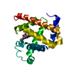
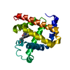
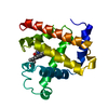
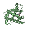
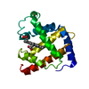
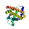
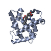
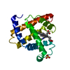
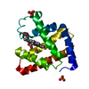
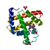
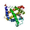
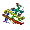
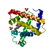
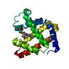
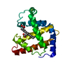
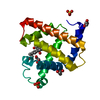
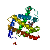
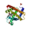
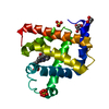
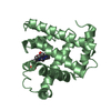
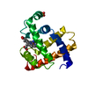
- PDB-1dtm: CRYSTAL STRUCTURE OF THE SPERM-WHALE MYOGLOBIN MUTANT H93G COMPLE... -
+ Open data

- Basic information
| Entry | Database: PDB / ID: 1dtm | ||||||
|---|---|---|---|---|---|---|---|
| Title | CRYSTAL STRUCTURE OF THE SPERM-WHALE MYOGLOBIN MUTANT H93G COMPLEXED WITH 4-METHYLIMIDAZOLE, METAQUO FORM | ||||||
 Components Components | RECOMBINANT SPERM WHALE MYOGLOBIN VARIANT H93G | ||||||
 Keywords Keywords | OXYGEN STORAGE/TRANSPORT / Heme protein / Myoglobin / ligand-substitution / OXYGEN STORAGE-TRANSPORT COMPLEX | ||||||
| Function / homology |  Function and homology information Function and homology informationOxidoreductases; Acting on other nitrogenous compounds as donors / nitrite reductase activity / sarcoplasm / Oxidoreductases; Acting on a peroxide as acceptor; Peroxidases / removal of superoxide radicals / oxygen carrier activity / peroxidase activity / oxygen binding / heme binding / extracellular exosome / metal ion binding Similarity search - Function | ||||||
| Biological species |  | ||||||
| Method |  X-RAY DIFFRACTION / Resolution: 2.13 Å X-RAY DIFFRACTION / Resolution: 2.13 Å | ||||||
 Authors Authors | Barrick, D. / Dahlquist, F.W. | ||||||
 Citation Citation | Journal: Proteins / Year: 2000 Title: Trans-substitution of the proximal hydrogen bond in myoglobin: I. Structural consequences of hydrogen bond deletion. Authors: Barrick, D. / Dahlquist, F.W. #1: Journal: To be PublishedTitle: Trans-substitution of the proximal hydrogen bond in myoglobin: II. Energetics, functional consequences, and implications for hemoglobin allostery Authors: Barrick, D. | ||||||
| History |
|
- Structure visualization
| Structure viewer | Molecule: MolmilJmol/JSmol |
|---|
- Downloads & links
-Download
| PDBx/mmCIF format | 1dtm.cif.gz | 44.6 KB | Display | PDBx/mmCIF format |
|---|---|---|---|---|
| PDB format | pdb1dtm.ent.gz | 30.7 KB | Display | PDB format |
| PDBx/mmJSON format | 1dtm.json.gz | Tree view | PDBx/mmJSON format | |
| Others |  Other downloads Other downloads |
-Validation report
| Arichive directory | https://data.pdbj.org/pub/pdb/validation_reports/dt/1dtmftp://data.pdbj.org/pub/pdb/validation_reports/dt/1dtm | HTTPS FTP |
|---|
-Related structure data


-Links
 PDBj
PDBj


- Assembly
| Deposited unit | 
| ||||||||
|---|---|---|---|---|---|---|---|---|---|
| 1 |
| ||||||||
| Unit cell |
|
-Components
| #1: Protein | Mass: 17153.857 Da / Num. of mol.: 1 / Mutation: H93G Source method: isolated from a genetically manipulated source Source: (gene. exp.)  |
|---|---|
| #2: Chemical | ChemComp-HEM /   Mass: 616.487 Da / Num. of mol.: 1 / Source method: obtained synthetically / Formula: C34H32FeN4O4 Mass: 616.487 Da / Num. of mol.: 1 / Source method: obtained synthetically / Formula: C34H32FeN4O4 |
| #3: Chemical | ChemComp-4MZ / 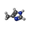  Mass: 82.104 Da / Num. of mol.: 1 / Source method: obtained synthetically / Formula: C4H6N2 Mass: 82.104 Da / Num. of mol.: 1 / Source method: obtained synthetically / Formula: C4H6N2Details: 4-methylimidazole from Aldrich Chemical Company (Cat # 19,988-5). 4-methylimidazole substituted for imidazole by extensive buffer exchange using spin-columns equilibrated with 0.1 M 4-methylimidazole. |
| #4: Water | ChemComp-HOH /  Mass: 18.015 Da / Num. of mol.: 15 / Source method: isolated from a natural source / Formula: H2O Mass: 18.015 Da / Num. of mol.: 15 / Source method: isolated from a natural source / Formula: H2O |
-Experimental details
-Experiment
| Experiment | Method: X-RAY DIFFRACTION / Number of used crystals: 1 |
|---|
- Sample preparation
| Crystal | Density Matthews: 2.26 Å3/Da / Density % sol: 45.69 % | ||||||||||||||||||||||||||||||||||||
|---|---|---|---|---|---|---|---|---|---|---|---|---|---|---|---|---|---|---|---|---|---|---|---|---|---|---|---|---|---|---|---|---|---|---|---|---|---|
| Crystal grow | Method: vapor diffusion, hanging drop / pH: 6.5 Details: 35 % PEG 8000, 0.3-0.35 M NaOAc, 0.1 M PIPES, and 0.1 % dioxane, 0.1 M 4-methylimidazole, pH 6.5, VAPOR DIFFUSION, HANGING DROP Temp details: Room temperature | ||||||||||||||||||||||||||||||||||||
| Crystal grow | *PLUS Details: drop consists of equal volume of protein and reservoir solutions | ||||||||||||||||||||||||||||||||||||
| Components of the solutions | *PLUS
|
-Data collection
| Diffraction | Mean temperature: 298 K |
|---|---|
| Diffraction source | Source: ROTATING ANODE / Type: RIGAKU RU200 / Wavelength: 1.54 |
| Detector | Type: SDMS / Detector: AREA DETECTOR / Date: Jul 29, 1994 |
| Radiation | Protocol: SINGLE WAVELENGTH / Monochromatic (M) / Laue (L): M / Scattering type: x-ray |
| Radiation wavelength | Wavelength: 1.54 Å / Relative weight: 1 |
| Reflection | Resolution: 2.13→20 Å / Num. all: 7356 / Num. obs: 7356 / % possible obs: 78.9 % / Redundancy: 2.63 % / Biso Wilson estimate: 23.3 Å2 / Rmerge(I) obs: 0.036 / Net I/σ(I): 12.54 |
| Reflection shell | Resolution: 2.13→2.298 Å / Redundancy: 1.51 % / Rmerge(I) obs: 0.141 / Num. unique all: 853 / % possible all: 49 |
| Reflection shell | *PLUS % possible obs: 49 % |
- Processing
| Software |
| ||||||||||||
|---|---|---|---|---|---|---|---|---|---|---|---|---|---|
| Refinement | Resolution: 2.13→20 Å Stereochemistry target values: Bond length rmsd 0.02 angstroms, Bond angle 3.0 degrees, B-correlation 6 square angstroms, Trigonal planarity 0.02, Planar groups 0.02 Details: 4-methyl imidazole built into an omit map in the proximal pocket after extensive all-atom refinement of the protein and heme
| ||||||||||||
| Refinement step | Cycle: LAST / Resolution: 2.13→20 Å
| ||||||||||||
| Refine LS restraints |
|
