 Movie
Movie Controller


+ Open data

- Basic information
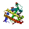
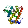
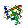
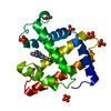
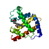
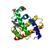
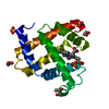
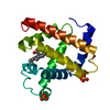
| Entry | Database: PDB / ID: 1a6m | ||||||
|---|---|---|---|---|---|---|---|
| Title | OXY-MYOGLOBIN, ATOMIC RESOLUTION | ||||||
 Components Components | MYOGLOBIN | ||||||
 Keywords Keywords | OXYGEN TRANSPORT / HEME PROTEIN / MODEL COMPOUNDS / OXYGEN STORAGE / LIGAND BINDING GEOMETRY / CONFORMATIONAL SUBSTATES | ||||||
| Function / homology |  Function and homology information Function and homology informationOxidoreductases; Acting on other nitrogenous compounds as donors / nitrite reductase activity / sarcoplasm / Oxidoreductases; Acting on a peroxide as acceptor; Peroxidases / removal of superoxide radicals / oxygen carrier activity / peroxidase activity / oxygen binding / heme binding / extracellular exosome / metal ion binding Similarity search - Function | ||||||
| Biological species |  | ||||||
| Method |  X-RAY DIFFRACTION / SYNCHROTRON / MOLECULAR REPLACEMENT / Resolution: 1 Å X-RAY DIFFRACTION / SYNCHROTRON / MOLECULAR REPLACEMENT / Resolution: 1 Å | ||||||
 Authors Authors | Vojtechovsky, J. / Chu, K. / Berendzen, J. / Sweet, R.M. / Schlichting, I. | ||||||
 Citation Citation | Journal: Biophys.J. / Year: 1999 Title: Crystal structures of myoglobin-ligand complexes at near-atomic resolution. Authors: Vojtechovsky, J. / Chu, K. / Berendzen, J. / Sweet, R.M. / Schlichting, I. | ||||||
| History |
|
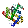
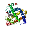
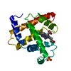
- Structure visualization
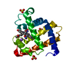
| Structure viewer | Molecule: MolmilJmol/JSmol |
|---|
- Downloads & links
-Download
| PDBx/mmCIF format | 1a6m.cif.gz | 89 KB | Display | PDBx/mmCIF format |
|---|---|---|---|---|
| PDB format | pdb1a6m.ent.gz | 67.6 KB | Display | PDB format |
| PDBx/mmJSON format | 1a6m.json.gz | Tree view | PDBx/mmJSON format | |
| Others |  Other downloads Other downloads |
-Validation report
| Arichive directory | https://data.pdbj.org/pub/pdb/validation_reports/a6/1a6mftp://data.pdbj.org/pub/pdb/validation_reports/a6/1a6m | HTTPS FTP |
|---|
-Related structure data
| Related structure data |  1a6gC 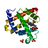 1a6kC  1a6nC  1mbcS S: Starting model for refinement C: citing same article ( |
|---|---|
| Similar structure data |


-Links
 PDBj
PDBj


- Assembly
| Deposited unit | 
| ||||||||
|---|---|---|---|---|---|---|---|---|---|
| 1 |
| ||||||||
| Unit cell |
|
-Components
| #1: Protein | Mass: 17049.771 Da / Num. of mol.: 1 / Source method: isolated from a natural source / Source: (natural) | ||||||
|---|---|---|---|---|---|---|---|
| #2: Chemical |   Mass: 96.063 Da / Num. of mol.: 2 / Source method: obtained synthetically / Formula: SO4 Mass: 96.063 Da / Num. of mol.: 2 / Source method: obtained synthetically / Formula: SO4#3: Chemical | ChemComp-HEM / |   Mass: 616.487 Da / Num. of mol.: 1 / Source method: obtained synthetically / Formula: C34H32FeN4O4 Mass: 616.487 Da / Num. of mol.: 1 / Source method: obtained synthetically / Formula: C34H32FeN4O4#4: Chemical | ChemComp-OXY / |   Mass: 31.999 Da / Num. of mol.: 1 / Source method: obtained synthetically / Formula: O2 Mass: 31.999 Da / Num. of mol.: 1 / Source method: obtained synthetically / Formula: O2#5: Water | ChemComp-HOH / |  Mass: 18.015 Da / Num. of mol.: 186 / Source method: isolated from a natural source / Formula: H2O Mass: 18.015 Da / Num. of mol.: 186 / Source method: isolated from a natural source / Formula: H2O |
-Experimental details
-Experiment
| Experiment | Method: X-RAY DIFFRACTION / Number of used crystals: 1 |
|---|
- Sample preparation
| Crystal | Density Matthews: 1.9 Å3/Da / Density % sol: 36 % | ||||||||||||||||||||
|---|---|---|---|---|---|---|---|---|---|---|---|---|---|---|---|---|---|---|---|---|---|
| Crystal grow | pH: 7 / Details: pH 7.0 | ||||||||||||||||||||
| Crystal grow | *PLUS Method: batch method | ||||||||||||||||||||
| Components of the solutions | *PLUS
|
-Data collection
| Diffraction | Mean temperature: 100 K |
|---|---|
| Diffraction source | Source: SYNCHROTRON / Site: NSLS  / Beamline: X8C / Wavelength: 0.84 / Beamline: X8C / Wavelength: 0.84 |
| Detector | Type: MARRESEARCH / Detector: IMAGE PLATE / Date: Jan 1, 1997 |
| Radiation | Monochromatic (M) / Laue (L): M / Scattering type: x-ray |
| Radiation wavelength | Wavelength: 0.84 Å / Relative weight: 1 |
| Reflection | Resolution: 1→50 Å / Num. obs: 67676 / % possible obs: 97 % / Observed criterion σ(I): -3 / Rsym value: 0.057 / Net I/σ(I): 26 |
| Reflection shell | Resolution: 1→1.1 Å / Mean I/σ(I) obs: 5 / Rsym value: 0.232 / % possible all: 88 |
| Reflection | *PLUS Num. measured all: 530931 / Rmerge(I) obs: 0.057 |
| Reflection shell | *PLUS % possible obs: 88 % / Rmerge(I) obs: 0.232 |
- Processing
| Software |
| |||||||||||||||||||||||||||||||||
|---|---|---|---|---|---|---|---|---|---|---|---|---|---|---|---|---|---|---|---|---|---|---|---|---|---|---|---|---|---|---|---|---|---|---|
| Refinement | Method to determine structure: MOLECULAR REPLACEMENT Starting model: 1MBC Resolution: 1→8 Å / Num. parameters: 14251 / Num. restraintsaints: 18357 / Cross valid method: FREE R-VALUE StereochEM target val spec case: HEME - PARAMETERS BASED ON CSD Stereochemistry target values: ENGH & HUBER Details: NO GEOMETRIC RESTRAINTS APPLIED TO IRON AND THE PLANAR ATOMS OF THE HEME. BAYESIAN WEIGHTING WAS APPLIED TO THE REFLECTION SET AT THE FINAL ROUND OF REFINEMENT. SEE TERWILLIGER AND ...Details: NO GEOMETRIC RESTRAINTS APPLIED TO IRON AND THE PLANAR ATOMS OF THE HEME. BAYESIAN WEIGHTING WAS APPLIED TO THE REFLECTION SET AT THE FINAL ROUND OF REFINEMENT. SEE TERWILLIGER AND BERENDZEN, ACTA CRYST. D52:743-748 (1996). THE SOLVENT MOLECULES 129 AND 139 CAN BE MODELED ONLY FOR ONE ALTERNATIVE PROTEIN CONFORMATION.
| |||||||||||||||||||||||||||||||||
| Solvent computation | Solvent model: MOEWS & KRETSINGER | |||||||||||||||||||||||||||||||||
| Refine analyze | Num. disordered residues: 26 / Occupancy sum hydrogen: 1263 / Occupancy sum non hydrogen: 1426.8 | |||||||||||||||||||||||||||||||||
| Refinement step | Cycle: LAST / Resolution: 1→8 Å
| |||||||||||||||||||||||||||||||||
| Refine LS restraints |
|
