 ムービー
ムービー コントローラー
 構造ビューア
 EMN検索について


-検索条件
-検索結果
検索 (著者・登録者: sengupta & u)の結果154件中、1から50件目までを表示しています

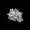
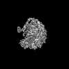
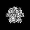
EMDB-54547: 
Cerebellar GluA1/4 NTD tetramer (focused refinement)

PDB-9s3q:
Cerebellar GluA1/4 NTD tetramer (focused refinement)

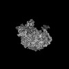
EMDB-54556:
Cerebellar GluA1/4 LBD tetramer (focused refinement)

PDB-9s3z:
Cerebellar GluA1/4 LBD tetramer (focused refinement)

EMDB-54543:
Cerebellar GluA2/4 NTD heterophilic tetramer interface (focused refinement)

EMDB-54558:
Cerebellar GluA1/4 TMD with TARP gamma 7 (focused refinement)

EMDB-54559:
Cerebellar GluAx/A4 TMD with four TARPs (focused refinement)

EMDB-55413:
GluA4 N-terminal domain bound to nanobody NB74 (focused refinement)

EMDB-55414:
GluA4 LBD-TMD with TARP gamma 2 (focused refinement)

EMDB-55418:
GluA4 with TARP gamma2 (consensus refinement)

EMDB-55419:
Full-length GluA4 with TARP gamma 2 (composite map)

PDB-9s3o:
Cerebellar GluA2/4 NTD heterophilic tetramer interface (focused refinement)

PDB-9s41:
Cerebellar GluA1/4 TMD with TARP gamma 7 (focused refinement)

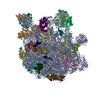
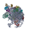
EMDB-61605:
50S Ribosomal Subunit precursor state III

EMDB-61613:
50S subunit precursor state I

EMDB-61625:
50S precursor - Erm complex (C-II)

EMDB-61781:
50S precursor - Erm complex (C-I)

PDB-9jmk:
50S Ribosomal Subunit precursor state III

PDB-9jns:
50S precursor - Erm complex (C-II)

PDB-9jsr:
50S precursor - Erm complex (C-I)

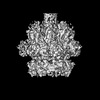
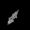
EMDB-45896:
cryo-EM structure of the Nipah virus polymerase containing the connecting domain

EMDB-48649:
Cryo-EM structure of a truncated Nipah virus (Malaysia Strain) L:P complex

PDB-9muw:
Cryo-EM structure of a truncated Nipah virus (Malaysia Strain) L:P complex

PDB-9mzh:
Cryo-EM structure of the Nipah virus polymerase containing the connecting domain

EMDB-45782:
Cryo-EM structure of the Nipah virus (Malaysia Strain) L:P complex

PDB-9cok:
Cryo-EM structure of the Nipah virus (Malaysia Strain) L:P complex

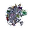
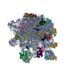
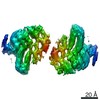
EMDB-61959:
EF-G2 bound 70S ribosome complex of M. smegmatis

EMDB-61960:
EF-G2 bound 50S ribosome subunit complex of M. smegmatis

PDB-9k0z:
EF-G2 bound 70S ribosome complex of M. smegmatis

PDB-9k10:
EF-G2 bound 50S ribosome subunit complex of M. smegmatis

EMDB-39462:
Cryo-EM map of 30S ribosomal subunit in complex with MetAP1c of Mycobacterium smegmatis

PDB-8yp6:
Cryo-EM map of 30S ribosomal subunit in complex with MetAP1c of Mycobacterium smegmatis
EMDB-37007:
Mycobacterium smegmatis 50S ribosomal subunit-HflX complex
EMDB-38788:
Mycobacterium smegmatis 50S ribosomal subunit with Erythromycin
PDB-8kab:
Mycobacterium smegmatis 50S ribosomal subunit-HflX complex
PDB-8xz3:
Mycobacterium smegmatis 50S ribosomal subunit with Erythromycin

EMDB-33096:
Mycobacterium smegmatis 50S ribosomal subunit from Stationary phase of growth
EMDB-33599:
Mycobacterium smegmatis 50S ribosomal subunit from Log Phase of growth
PDB-7xam:
Mycobacterium smegmatis 50S ribosomal subunit from Stationary phase of growth
PDB-7y41:
Mycobacterium smegmatis 50S ribosomal subunit from Log Phase of growth

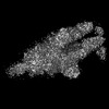
EMDB-33752:
Cryo-EM structure of Mycobacterial Type VII Secretion System Virulence Factor EspB (residues 1-332) with Phosphatidic acid (PA)

EMDB-34878:
Cryo-EM structure of Mycobacterial Type VII Secretion System Virulence Factor EspB (residues 1-332)

PDB-7yl9:
Cryo-EM structure of complete transmembrane channel E289A mutant Vibrio cholerae Cytolysin
EMDB-33215:
Cryo-EM reconstruction of complete transmembrane channel E289A mutant Vibrio cholerae Cytolysin
EMDB-33219:
Cryo-EM reconstruction of partial transmembrane channel E289A mutant Vibrio cholerae Cytolysin
EMDB-32388:
Cryo-EM 3D model of the 3-RBD up dimeric spike protein of SARS-CoV2 in the presence of SIH-5
EMDB-33042:
Cryo-EM 3D model of the 3-RBD up single trimeric spike protein of SARS-CoV2 in the presence of synthetic peptide SIH-5.

PDB-7x7n:
3D model of the 3-RBD up single trimeric spike protein of SARS-CoV2 in the presence of synthetic peptide SIH-5.
EMDB-23212:
Alpha-synuclein fibrils

EMDB-31972:
Cryo-EM 3D reconstruction of Vibrio cholerae Cytolysin embedded in lipid bilayer- State 3
ページ:
 wwPDBはEMDBデータモデルのバージョン3へ移行します
