Movie
Movie Controller


+ Open data

- Basic information
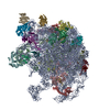
| Entry | Database: PDB / ID: 9k10 | |||||||||||||||||||||
|---|---|---|---|---|---|---|---|---|---|---|---|---|---|---|---|---|---|---|---|---|---|---|
| Title | EF-G2 bound 50S ribosome subunit complex of M. smegmatis | |||||||||||||||||||||
 Components Components |
| |||||||||||||||||||||
 Keywords Keywords | RIBOSOME / Mycobacterium 50S ribosomal subunit / elongation factor G2 / stationary phase | |||||||||||||||||||||
| Function / homology |  Function and homology information Function and homology informationribosome disassembly / translation elongation factor activity / large ribosomal subunit / transferase activity / 5S rRNA binding / ribosomal large subunit assembly / large ribosomal subunit rRNA binding / cytosolic large ribosomal subunit / cytoplasmic translation / tRNA binding ...ribosome disassembly / translation elongation factor activity / large ribosomal subunit / transferase activity / 5S rRNA binding / ribosomal large subunit assembly / large ribosomal subunit rRNA binding / cytosolic large ribosomal subunit / cytoplasmic translation / tRNA binding / negative regulation of translation / rRNA binding / structural constituent of ribosome / ribosome / translation / ribonucleoprotein complex / mRNA binding / GTPase activity / GTP binding / metal ion binding / cytoplasm Similarity search - Function | |||||||||||||||||||||
| Biological species |  Mycolicibacterium smegmatis MC2 155 (bacteria) Mycolicibacterium smegmatis MC2 155 (bacteria) | |||||||||||||||||||||
| Method | ELECTRON MICROSCOPY / single particle reconstruction / cryo EM / Resolution: 3.6 Å | |||||||||||||||||||||
 Authors Authors | Sengupta, J. / Baid, P. | |||||||||||||||||||||
| Funding support |  India, 3items India, 3items
| |||||||||||||||||||||
 Citation Citation | Journal: FEBS J / Year: 2025 Title: Cryo-EM structural analyses reveal a unique role for elongation factor G2 (EF-G2) in Mycobacteria. Authors: Priya Baid / Jayati Sengupta / Abstract: The gene-encoding translation elongation factor G (EF-G) has undergone gene duplication across various bacterial species including Mycobacteria, and in mammalian mitochondria, leading to the ...The gene-encoding translation elongation factor G (EF-G) has undergone gene duplication across various bacterial species including Mycobacteria, and in mammalian mitochondria, leading to the emergence of the paralogue elongation factor G2 (EF-G2). Our study reveals that mycobacterial EF-G2, unlike EF-G1, neither participates in ribosome-recycling nor significantly contributes to overall translation, suggesting that it plays an alternative role in Mycobacteria. Remarkably, our investigation found a significant overexpression of mycobacterial EF-G2 during the stationary growth phase. Moreover, EF-G2 lacks ribosome-dependent GTPase activity, an observation consistent with previous reports. Cryo-EM analysis of the M. smegmatis 70S ribosome purified from the nutrient-starved (stationary) phase and complexed with EF-G2 unveiled the structural basis for its inability to hydrolyse GTP in a ribosome-dependent manner. Furthermore, we report an unprecedented binding mode of two EF-G2 copies on the 50S ribosomal subunit that impedes subunit association, thereby preventing the formation of active 70S ribosomes. Thus, instead of performing canonical functions, mycobacterial EF-G2 acts as a translation repressor during nutrient starvation. Altogether, our findings shed light on the multifaceted mechanisms by which EF-G2 modulates protein synthesis under nutrient-limited conditions, providing insights into adaptive strategies employed by Mycobacteria to survive in hostile environments. | |||||||||||||||||||||
| History |
|
- Structure visualization
| Structure viewer | Molecule: MolmilJmol/JSmol |
|---|
- Downloads & links
-Download
| PDBx/mmCIF format | 9k10.cif.gz | 2.4 MB | Display | PDBx/mmCIF format |
|---|---|---|---|---|
| PDB format | pdb9k10.ent.gz | Display | PDB format | |
| PDBx/mmJSON format | 9k10.json.gz | Tree view | PDBx/mmJSON format | |
| Others |  Other downloads Other downloads |
-Validation report
| Arichive directory | https://data.pdbj.org/pub/pdb/validation_reports/k1/9k10ftp://data.pdbj.org/pub/pdb/validation_reports/k1/9k10 | HTTPS FTP |
|---|
-Related structure data
| Related structure data |  61960MC  9k0zC M: map data used to model this data C: citing same article ( |
|---|---|
| Similar structure data |

-Links
 PDBj
PDBj


- Assembly
| Deposited unit | 
|
|---|---|
| 1 |
|
-Components
+50S ribosomal protein ... , 31 types, 31 molecules 3CDEFGHIJKLMNOPQRSTUWXYZabcdefg
-Protein , 2 types, 3 molecules 56V
| #2: Protein | Mass: 75420.797 Da / Num. of mol.: 2 Source method: isolated from a genetically manipulated source Source: (gene. exp.) Mycolicibacterium smegmatis MC2 155 (bacteria)Gene: MSMEG_6535 / Production host: #23: Protein | | Mass: 11228.946 Da / Num. of mol.: 1 / Source method: isolated from a natural source Source: (natural) Mycolicibacterium smegmatis MC2 155 (bacteria)References: UniProt: A0QSG0 |
|---|
-RNA chain , 2 types, 2 molecules BA
| #3: RNA chain | Mass: 38061.816 Da / Num. of mol.: 1 / Source method: isolated from a natural source Source: (natural) Mycolicibacterium smegmatis MC2 155 (bacteria)References: GenBank: 118168627 |
|---|---|
| #35: RNA chain | Mass: 1014391.625 Da / Num. of mol.: 1 / Source method: isolated from a natural source Source: (natural) Mycolicibacterium smegmatis MC2 155 (bacteria) |
-Non-polymers , 3 types, 413 molecules 


| #36: Chemical |  Mass: 522.196 Da / Num. of mol.: 2 / Source method: obtained synthetically / Formula: C10H17N6O13P3 / Feature type: SUBJECT OF INVESTIGATION Mass: 522.196 Da / Num. of mol.: 2 / Source method: obtained synthetically / Formula: C10H17N6O13P3 / Feature type: SUBJECT OF INVESTIGATIONComment: GppNHp, GMPPNP, energy-carrying molecule analogue*YM #37: Chemical | ChemComp-MG /  Mass: 24.305 Da / Num. of mol.: 407 / Source method: obtained synthetically / Formula: Mg Mass: 24.305 Da / Num. of mol.: 407 / Source method: obtained synthetically / Formula: Mg#38: Chemical | ChemComp-ZN /  Mass: 65.409 Da / Num. of mol.: 4 / Source method: obtained synthetically / Formula: Zn Mass: 65.409 Da / Num. of mol.: 4 / Source method: obtained synthetically / Formula: Zn |
|---|
-Details
| Has ligand of interest | Y |
|---|---|
| Has protein modification | N |
-Experimental details
-Experiment
| Experiment | Method: ELECTRON MICROSCOPY |
|---|---|
| EM experiment | Aggregation state: PARTICLE / 3D reconstruction method: single particle reconstruction |
- Sample preparation
| Component | Name: EF-G2 bound 50S ribosomal subunit complex of M. smegmatis Type: COMPLEX / Entity ID: #1-#2, #35, #3-#34 / Source: NATURAL |
|---|---|
| Molecular weight | Value: 1.8 MDa / Experimental value: YES |
| Source (natural) | Organism: Mycolicibacterium smegmatis MC2 155 (bacteria) |
| Buffer solution | pH: 7.8 |
| Specimen | Embedding applied: NO / Shadowing applied: NO / Staining applied: NO / Vitrification applied: YES |
| Specimen support | Grid material: COPPER / Grid mesh size: 300 divisions/in. / Grid type: Quantifoil R2/2 |
| Vitrification | Instrument: FEI VITROBOT MARK IV / Cryogen name: ETHANE / Humidity: 100 % |
- Electron microscopy imaging
| Experimental equipment |  Model: Titan Krios / Image courtesy: FEI Company |
|---|---|
| Microscopy | Model: TFS KRIOS |
| Electron gun | Electron source:  FIELD EMISSION GUN / Accelerating voltage: 300 kV / Illumination mode: FLOOD BEAM FIELD EMISSION GUN / Accelerating voltage: 300 kV / Illumination mode: FLOOD BEAM |
| Electron lens | Mode: BRIGHT FIELD / Nominal defocus max: 3300 nm / Nominal defocus min: 1800 nm / Cs: 2.7 mm |
| Specimen holder | Specimen holder model: FEI TITAN KRIOS AUTOGRID HOLDER |
| Image recording | Electron dose: 54 e/Å2 / Film or detector model: FEI FALCON III (4k x 4k) |
- Processing
| EM software | Name: PHENIX / Version: 1.20.1_4487: / Category: model refinement | ||||||||||||||||||||||||
|---|---|---|---|---|---|---|---|---|---|---|---|---|---|---|---|---|---|---|---|---|---|---|---|---|---|
| CTF correction | Type: PHASE FLIPPING AND AMPLITUDE CORRECTION | ||||||||||||||||||||||||
| Symmetry | Point symmetry: C1 (asymmetric) | ||||||||||||||||||||||||
| 3D reconstruction | Resolution: 3.6 Å / Resolution method: FSC 0.143 CUT-OFF / Num. of particles: 39621 / Symmetry type: POINT | ||||||||||||||||||||||||
| Atomic model building | Protocol: FLEXIBLE FIT / Space: REAL | ||||||||||||||||||||||||
| Atomic model building | PDB-ID: 7XAM Accession code: 7XAM / Source name: PDB / Type: experimental model | ||||||||||||||||||||||||
| Refine LS restraints |
|