Movie
Movie Controller

[English] 日本語
 Yorodumi
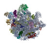
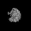
- PDB-7xam: Mycobacterium smegmatis 50S ribosomal subunit from Stationary pha... -
+ Open data

- Basic information
| Entry | Database: PDB / ID: 7xam | |||||||||||||||
|---|---|---|---|---|---|---|---|---|---|---|---|---|---|---|---|---|
| Title | Mycobacterium smegmatis 50S ribosomal subunit from Stationary phase of growth | |||||||||||||||
 Components Components |
| |||||||||||||||
 Keywords Keywords | RIBOSOME / 50S Subunit / Domain IV of 23S rRNA / Alternate conformation / Helix 68 | |||||||||||||||
| Function / homology |  Function and homology information Function and homology informationlarge ribosomal subunit / transferase activity / 5S rRNA binding / ribosomal large subunit assembly / large ribosomal subunit rRNA binding / cytosolic large ribosomal subunit / cytoplasmic translation / tRNA binding / negative regulation of translation / rRNA binding ...large ribosomal subunit / transferase activity / 5S rRNA binding / ribosomal large subunit assembly / large ribosomal subunit rRNA binding / cytosolic large ribosomal subunit / cytoplasmic translation / tRNA binding / negative regulation of translation / rRNA binding / structural constituent of ribosome / ribosome / translation / ribonucleoprotein complex / mRNA binding / metal ion binding / cytoplasm Similarity search - Function | |||||||||||||||
| Biological species |  Mycolicibacterium smegmatis MC2 155 (bacteria) Mycolicibacterium smegmatis MC2 155 (bacteria) | |||||||||||||||
| Method | ELECTRON MICROSCOPY / single particle reconstruction / cryo EM / Resolution: 3.5 Å | |||||||||||||||
 Authors Authors | Sengupta, J. / Baid, P. | |||||||||||||||
| Funding support |  India, 4items India, 4items
| |||||||||||||||
 Citation Citation | Journal: Int J Biol Macromol / Year: 2023 Title: Cryo-EM captures a unique conformational rearrangement in 23S rRNA helices of the Mycobacterium 50S subunit. Authors: Priya Baid / Jayati Sengupta / Abstract: Structural investigations of the ribosomes isolated from pathogenic and non-pathogenic Mycobacterium species have identified several mycobacteria-specific structural features of ribosomal RNA and ...Structural investigations of the ribosomes isolated from pathogenic and non-pathogenic Mycobacterium species have identified several mycobacteria-specific structural features of ribosomal RNA and proteins. Here, we report structural evidence of a hitherto unknown conformational switch of mycobacterium 23S rRNA helices (H54a and H67-H71). Cryo-electron microscopy (cryo-EM) structures (~3-4 Å) of the M. smegmatis (Msm) log-phase 50S ribosomal subunit revealed conformational variability in H67-H71 region of the 23S rRNA, and manifested that, while H68 possesses the usual stretched conformation in one class of the maps, another one exhibits a bulge-out, fused density of H68-H69 at the inter-subunit surface, indicating an intrinsic dynamics of these rRNA helices. Remarkably, altered conformation of H68 forming a more prominent bulge-out structure at the inter-subunit surface of the 50S subunit due to the conformational rearrangements of 23S rRNA H67-H71 region was clearly visualized in a 3 Å cryo-EM map of the 50S subunit obtained from the stationary phase ribosome dataset. The Msm50S subunit having such bulge-out conformation at the intersubunit surface would be incompatible for associating with the 30S subunit due to its inability to form major inter-subunit bridges. Evidently, availability of active 70S ribosome pool can be modulated by stabilizing either one of the H68 conformation. | |||||||||||||||
| History |
|
- Structure visualization
| Structure viewer | Molecule: MolmilJmol/JSmol |
|---|
- Downloads & links
-Download
| PDBx/mmCIF format | 7xam.cif.gz | 2.1 MB | Display | PDBx/mmCIF format |
|---|---|---|---|---|
| PDB format | pdb7xam.ent.gz | 1.7 MB | Display | PDB format |
| PDBx/mmJSON format | 7xam.json.gz | Tree view | PDBx/mmJSON format | |
| Others |  Other downloads Other downloads |
-Validation report
| Arichive directory | https://data.pdbj.org/pub/pdb/validation_reports/xa/7xamftp://data.pdbj.org/pub/pdb/validation_reports/xa/7xam | HTTPS FTP |
|---|
-Related structure data
| Related structure data |  33096MC 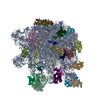 7y41C M: map data used to model this data C: citing same article ( |
|---|---|
| Similar structure data |
-Links
 PDBj
PDBj


- Assembly
| Deposited unit | 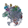
|
|---|---|
| 1 |
|
-Components
+50S ribosomal protein ... , 32 types, 32 molecules 3CDEFGHIJKLMNOPQRSTUVWXYZabcdefg
-RNA chain , 2 types, 2 molecules AB
| #2: RNA chain | Mass: 1012140.938 Da / Num. of mol.: 1 / Source method: isolated from a natural source Source: (natural) Mycolicibacterium smegmatis MC2 155 (bacteria)References: GenBank: 118168627 |
|---|---|
| #3: RNA chain | Mass: 38061.816 Da / Num. of mol.: 1 / Source method: isolated from a natural source Source: (natural) Mycolicibacterium smegmatis MC2 155 (bacteria)References: GenBank: 118168627 |
-Non-polymers , 2 types, 411 molecules 

| #35: Chemical | ChemComp-MG /  Mass: 24.305 Da / Num. of mol.: 407 / Source method: obtained synthetically / Formula: Mg / Feature type: SUBJECT OF INVESTIGATION Mass: 24.305 Da / Num. of mol.: 407 / Source method: obtained synthetically / Formula: Mg / Feature type: SUBJECT OF INVESTIGATION#36: Chemical | ChemComp-ZN /  Mass: 65.409 Da / Num. of mol.: 4 / Source method: obtained synthetically / Formula: Zn / Feature type: SUBJECT OF INVESTIGATION Mass: 65.409 Da / Num. of mol.: 4 / Source method: obtained synthetically / Formula: Zn / Feature type: SUBJECT OF INVESTIGATION |
|---|
-Details
| Has ligand of interest | Y |
|---|
-Experimental details
-Experiment
| Experiment | Method: ELECTRON MICROSCOPY |
|---|---|
| EM experiment | Aggregation state: PARTICLE / 3D reconstruction method: single particle reconstruction |
- Sample preparation
| Component | Name: Mycobacterium smegmatis 50S ribosomal subunit from Stationary Phase of growth Type: RIBOSOME / Entity ID: #1-#34 / Source: NATURAL |
|---|---|
| Molecular weight | Value: 1.8 MDa / Experimental value: YES |
| Source (natural) | Organism: Mycolicibacterium smegmatis MC2 155 (bacteria) |
| Buffer solution | pH: 7.8 |
| Specimen | Embedding applied: NO / Shadowing applied: NO / Staining applied: NO / Vitrification applied: YES |
| Specimen support | Grid material: COPPER / Grid mesh size: 300 divisions/in. / Grid type: Quantifoil R2/2 |
| Vitrification | Instrument: FEI VITROBOT MARK IV / Cryogen name: ETHANE / Humidity: 100 % |
- Electron microscopy imaging
| Experimental equipment |  Model: Titan Krios / Image courtesy: FEI Company |
|---|---|
| Microscopy | Model: TFS KRIOS |
| Electron gun | Electron source:  FIELD EMISSION GUN / Accelerating voltage: 300 kV / Illumination mode: FLOOD BEAM FIELD EMISSION GUN / Accelerating voltage: 300 kV / Illumination mode: FLOOD BEAM |
| Electron lens | Mode: BRIGHT FIELD / Nominal defocus max: 3300 nm / Nominal defocus min: 1800 nm / Cs: 2.7 mm |
| Specimen holder | Specimen holder model: FEI TITAN KRIOS AUTOGRID HOLDER |
| Image recording | Electron dose: 54 e/Å2 / Film or detector model: FEI FALCON III (4k x 4k) |
- Processing
| EM software |
| ||||||||||||||||||||||||||||||||||||
|---|---|---|---|---|---|---|---|---|---|---|---|---|---|---|---|---|---|---|---|---|---|---|---|---|---|---|---|---|---|---|---|---|---|---|---|---|---|
| CTF correction | Type: PHASE FLIPPING AND AMPLITUDE CORRECTION | ||||||||||||||||||||||||||||||||||||
| Symmetry | Point symmetry: C1 (asymmetric) | ||||||||||||||||||||||||||||||||||||
| 3D reconstruction | Resolution: 3.5 Å / Resolution method: FSC 0.143 CUT-OFF / Num. of particles: 172541 / Num. of class averages: 9 / Symmetry type: POINT | ||||||||||||||||||||||||||||||||||||
| Atomic model building | Protocol: FLEXIBLE FIT / Space: REAL | ||||||||||||||||||||||||||||||||||||
| Atomic model building | PDB-ID: 5O60 Accession code: 5O60 / Source name: PDB / Type: experimental model | ||||||||||||||||||||||||||||||||||||
| Refinement | Highest resolution: 3.5 Å |