- EMDB-33599: Mycobacterium smegmatis 50S ribosomal subunit from Log Phase of growth -
+
Open data
ID or keywords:
Loading...
-
Basic information
Entry
Database: EMDB / ID: EMD-33599
Title
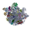
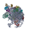
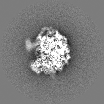
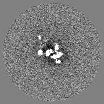
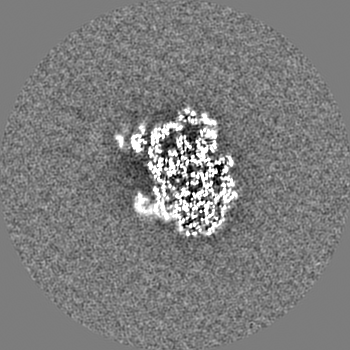
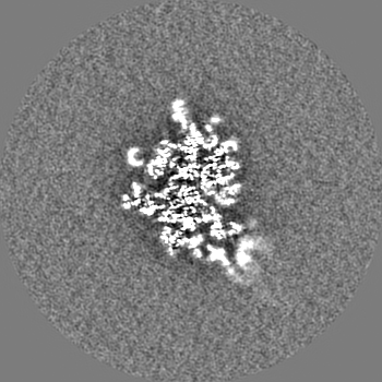
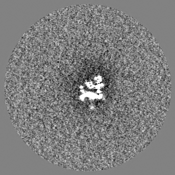
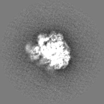
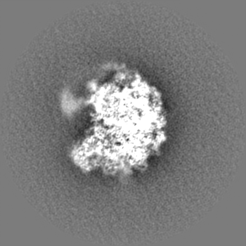
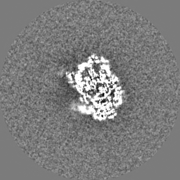
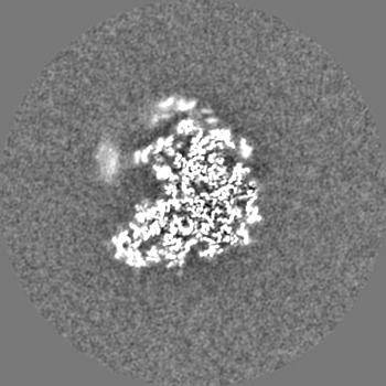
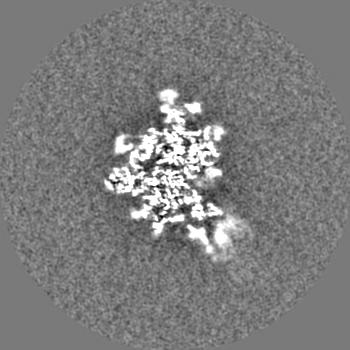
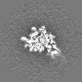
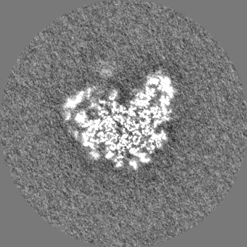
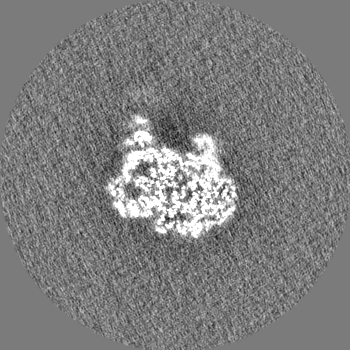
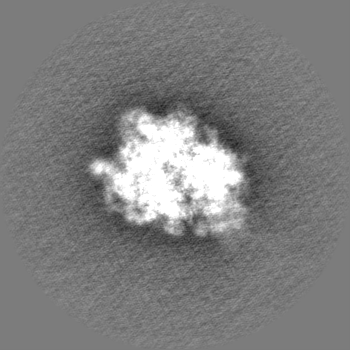
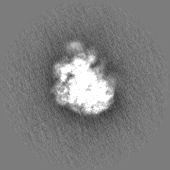
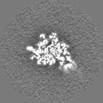
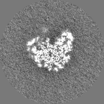
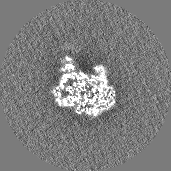
Mycobacterium smegmatis 50S ribosomal subunit from Log Phase of growth
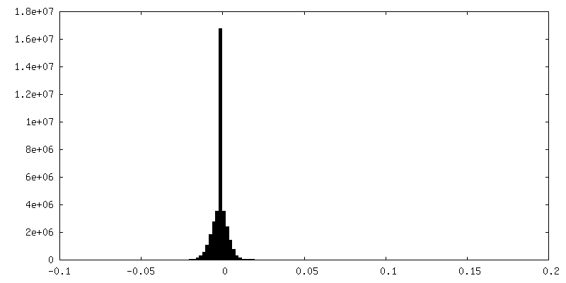
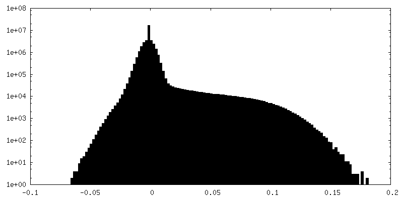
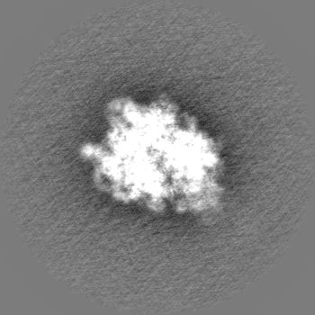
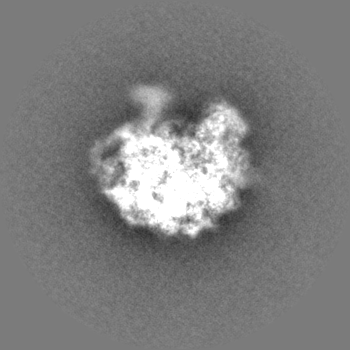
Map data
Mycobacterium smegmatis 50S ribosomal subunit from Log Phase of growth
Sample
Complex: Mycobacterium smegmatis 50S ribosomal subunit from Log Phase of growth
Protein or peptide: x 31 types
RNA: x 2 types
Ligand: x 2 types
Keywords
Mycobacterium 50S subunit / Domain IV of 23S rRNA / Log Phase / RIBOSOME
Function / homology
Function and homology information
large ribosomal subunit / transferase activity / 5S rRNA binding / ribosomal large subunit assembly / large ribosomal subunit rRNA binding / cytosolic large ribosomal subunit / cytoplasmic translation / tRNA binding / negative regulation of translation / rRNA binding ...large ribosomal subunit / transferase activity / 5S rRNA binding / ribosomal large subunit assembly / large ribosomal subunit rRNA binding / cytosolic large ribosomal subunit / cytoplasmic translation / tRNA binding / negative regulation of translation / rRNA binding / structural constituent of ribosome / ribosome / translation / ribonucleoprotein complex / mRNA binding / metal ion binding / cytoplasm Similarity search - Function
: / 50S ribosomal protein L37 helical region / Ribosomal protein L10, eubacterial, conserved site / Ribosomal protein L10 signature. / Ribosomal protein L10 / Ribosomal protein L25, long-form / Ribosomal protein L25, beta domain / Ribosomal protein L25, C-terminal / Ribosomal protein TL5, C-terminal domain / : ...: / 50S ribosomal protein L37 helical region / Ribosomal protein L10, eubacterial, conserved site / Ribosomal protein L10 signature. / Ribosomal protein L10 / Ribosomal protein L25, long-form / Ribosomal protein L25, beta domain / Ribosomal protein L25, C-terminal / Ribosomal protein TL5, C-terminal domain / : / : / Ribosomal protein L11, bacterial-type / Ribosomal protein L31 type A / Ribosomal protein L31 signature. / Ribosomal protein L11, conserved site / Ribosomal protein L11 signature. / Ribosomal protein L31 / Ribosomal protein L31 superfamily / Ribosomal protein L31 / Ribosomal protein L10-like domain superfamily / Ribosomal protein L10P / Ribosomal protein L10 / Ribosomal protein L16 signature 1. / Ribosomal protein L6, conserved site / Ribosomal protein L6 signature 1. / Ribosomal protein L21, conserved site / Ribosomal protein L21 signature. / : / Ribosomal protein L11, N-terminal / Ribosomal protein L11, N-terminal domain / Ribosomal protein L11/L12 / Ribosomal protein L11, C-terminal / Ribosomal protein L11, C-terminal domain superfamily / Ribosomal protein L11/L12, N-terminal domain superfamily / Ribosomal protein L11/L12 / Ribosomal protein L11, RNA binding domain / Ribosomal protein L16 signature 2. / Ribosomal protein L16, conserved site / Ribosomal protein L17 signature. / Ribosomal L25p family / Ribosomal protein L25 / Ribosomal protein L36 signature. / Ribosomal protein L25/Gln-tRNA synthetase, N-terminal / Ribosomal protein L25/Gln-tRNA synthetase, anti-codon-binding domain superfamily / : / Ribosomal protein L33, conserved site / Ribosomal protein L33 signature. / Ribosomal protein L28/L24 superfamily / Ribosomal protein L35, conserved site / Ribosomal protein L35 signature. / Ribosomal protein L28 / Ribosomal protein L35, non-mitochondrial / Ribosomal protein L18, bacterial-type / : / Ribosomal protein L6, bacterial-type / Ribosomal protein L5, bacterial-type / Ribosomal protein L19, conserved site / Ribosomal protein L19 signature. / : / Ribosomal protein L36 / Ribosomal protein L36 superfamily / Ribosomal protein L36 / Ribosomal protein L20 signature. / Ribosomal protein L34, conserved site / Ribosomal protein L34 signature. / Ribosomal protein L14P, bacterial-type / Ribosomal protein L27, conserved site / Ribosomal protein L27 signature. / Ribosomal protein L35 / Ribosomal protein L35 superfamily / Ribosomal protein L22, bacterial/chloroplast-type / Ribosomal protein L35 / Ribosomal protein L33 / Ribosomal protein L18 / Ribosomal L18 of archaea, bacteria, mitoch. and chloroplast / Ribosomal protein L33 / Ribosomal protein L2, bacterial/organellar-type / Ribosomal L28 family / Ribosomal protein L33 superfamily / Ribosomal protein L28/L24 / Ribosomal protein L30, bacterial-type / L28p-like / Ribosomal protein L16 / Ribosomal protein L20 / Ribosomal protein L20 / Ribosomal protein L20, C-terminal / Ribosomal protein L19 / Ribosomal protein L19 / Ribosomal protein L19 superfamily / : / Large ribosomal subunit protein uL24, C-terminal domain / Ribosomal protein L17 / Ribosomal protein L17 superfamily / Ribosomal protein L17 / Ribosomal protein L27 / Ribosomal L27 protein / Ribosomal protein L34 / Ribosomal protein L34 / Ribosomal protein L24 / Ribosomal L32p protein family Similarity search - Domain/homology
Large ribosomal subunit protein bL33A / Large ribosomal subunit protein uL11 / Large ribosomal subunit protein uL10 / Large ribosomal subunit protein uL3 / Large ribosomal subunit protein uL4 / Large ribosomal subunit protein uL23 / Large ribosomal subunit protein uL2 / Large ribosomal subunit protein uL22 / Large ribosomal subunit protein uL16 / Large ribosomal subunit protein uL29 ...Large ribosomal subunit protein bL33A / Large ribosomal subunit protein uL11 / Large ribosomal subunit protein uL10 / Large ribosomal subunit protein uL3 / Large ribosomal subunit protein uL4 / Large ribosomal subunit protein uL23 / Large ribosomal subunit protein uL2 / Large ribosomal subunit protein uL22 / Large ribosomal subunit protein uL16 / Large ribosomal subunit protein uL29 / Large ribosomal subunit protein uL14 / Large ribosomal subunit protein uL24 / Large ribosomal subunit protein uL5 / Large ribosomal subunit protein uL6 / Large ribosomal subunit protein uL18 / Large ribosomal subunit protein uL30 / Large ribosomal subunit protein uL15 / Large ribosomal subunit protein bL36 / Large ribosomal subunit protein bL17 / Large ribosomal subunit protein uL13 / Uncharacterized protein / Large ribosomal subunit protein bL28 / Large ribosomal subunit protein bL19 / Large ribosomal subunit protein bL20 / Large ribosomal subunit protein bL35 / Large ribosomal subunit protein bL27 / Large ribosomal subunit protein bL21 / Large ribosomal subunit protein bL31 / Large ribosomal subunit protein bL25 / Large ribosomal subunit protein bL32 / Large ribosomal subunit protein bL34 Similarity search - Component
Biological species
Mycolicibacterium smegmatis MC2 155 (bacteria)
Method
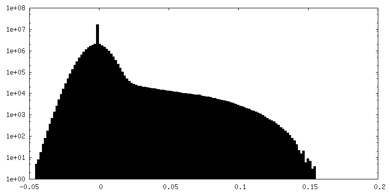
single particle reconstruction / cryo EM / Resolution: 4.1 Å
Council of Scientific & Industrial Research (CSIR)
MLP-139
India
Citation
Journal: Int J Biol Macromol / Year: 2023 Title: Cryo-EM captures a unique conformational rearrangement in 23S rRNA helices of the Mycobacterium 50S subunit. Authors: Priya Baid / Jayati Sengupta / Abstract: Structural investigations of the ribosomes isolated from pathogenic and non-pathogenic Mycobacterium species have identified several mycobacteria-specific structural features of ribosomal RNA and ...Structural investigations of the ribosomes isolated from pathogenic and non-pathogenic Mycobacterium species have identified several mycobacteria-specific structural features of ribosomal RNA and proteins. Here, we report structural evidence of a hitherto unknown conformational switch of mycobacterium 23S rRNA helices (H54a and H67-H71). Cryo-electron microscopy (cryo-EM) structures (~3-4 Å) of the M. smegmatis (Msm) log-phase 50S ribosomal subunit revealed conformational variability in H67-H71 region of the 23S rRNA, and manifested that, while H68 possesses the usual stretched conformation in one class of the maps, another one exhibits a bulge-out, fused density of H68-H69 at the inter-subunit surface, indicating an intrinsic dynamics of these rRNA helices. Remarkably, altered conformation of H68 forming a more prominent bulge-out structure at the inter-subunit surface of the 50S subunit due to the conformational rearrangements of 23S rRNA H67-H71 region was clearly visualized in a 3 Å cryo-EM map of the 50S subunit obtained from the stationary phase ribosome dataset. The Msm50S subunit having such bulge-out conformation at the intersubunit surface would be incompatible for associating with the 30S subunit due to its inability to form major inter-subunit bridges. Evidently, availability of active 70S ribosome pool can be modulated by stabilizing either one of the H68 conformation.
In the structure databanks used in Yorodumi, some data are registered as the other names, "COVID-19 virus" and "2019-nCoV". Here are the details of the virus and the list of structure data.
Jan 31, 2019. EMDB accession codes are about to change! (news from PDBe EMDB page)
EMDB accession codes are about to change! (news from PDBe EMDB page)
The allocation of 4 digits for EMDB accession codes will soon come to an end. Whilst these codes will remain in use, new EMDB accession codes will include an additional digit and will expand incrementally as the available range of codes is exhausted. The current 4-digit format prefixed with “EMD-” (i.e. EMD-XXXX) will advance to a 5-digit format (i.e. EMD-XXXXX), and so on. It is currently estimated that the 4-digit codes will be depleted around Spring 2019, at which point the 5-digit format will come into force.
The EM Navigator/Yorodumi systems omit the EMD- prefix.
Related info.:Q: What is EMD? / ID/Accession-code notation in Yorodumi/EM Navigator
Yorodumi is a browser for structure data from EMDB, PDB, SASBDB, etc.
This page is also the successor to EM Navigator detail page, and also detail information page/front-end page for Omokage search.
The word "yorodu" (or yorozu) is an old Japanese word meaning "ten thousand". "mi" (miru) is to see.
Related info.:EMDB / PDB / SASBDB / Comparison of 3 databanks / Yorodumi Search / Aug 31, 2016. New EM Navigator & Yorodumi / Yorodumi Papers / Jmol/JSmol / Function and homology information / Changes in new EM Navigator and Yorodumi
 Movie
Movie Controller

 Yorodumi
 Open data
 Basic information
 Map data
 Sample
 Keywords
 Function and homology information
 Mycolicibacterium smegmatis MC2 155 (bacteria)
 Authors
 India, 2 items
  Citation
 Structure visualization
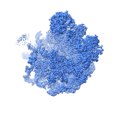
 Downloads & links
 emd_33599.png
 http://ftp.pdbj.org/pub/emdb/structures/EMD-33599
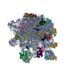


 X (Sec.)
 Y (Row.)
 Z (Col.)


 Sample components
 Processing
 Electron microscopy
 FIELD EMISSION GUN