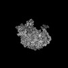
 Movie
Movie Controller


+ Open data

- Basic information
| Entry | Database: PDB / ID: 8kab | |||||||||
|---|---|---|---|---|---|---|---|---|---|---|
| Title | Mycobacterium smegmatis 50S ribosomal subunit-HflX complex | |||||||||
 Components Components |
| |||||||||
 Keywords Keywords | RIBOSOME / Complex | |||||||||
| Function / homology |  Function and homology information Function and homology informationlarge ribosomal subunit / transferase activity / ribosome binding / 5S rRNA binding / ribosomal large subunit assembly / large ribosomal subunit rRNA binding / cytosolic large ribosomal subunit / cytoplasmic translation / tRNA binding / negative regulation of translation ...large ribosomal subunit / transferase activity / ribosome binding / 5S rRNA binding / ribosomal large subunit assembly / large ribosomal subunit rRNA binding / cytosolic large ribosomal subunit / cytoplasmic translation / tRNA binding / negative regulation of translation / rRNA binding / structural constituent of ribosome / ribosome / translation / ribonucleoprotein complex / mRNA binding / GTPase activity / GTP binding / metal ion binding / cytoplasm Similarity search - Function | |||||||||
| Biological species |  Mycolicibacterium smegmatis MC2 155 (bacteria) Mycolicibacterium smegmatis MC2 155 (bacteria) | |||||||||
| Method | ELECTRON MICROSCOPY / single particle reconstruction / cryo EM / Resolution: 3.3 Å | |||||||||
 Authors Authors | Srinivasan, K. / Banerjee, A. / Sengupta, J. | |||||||||
| Funding support |  India, 2items India, 2items
| |||||||||
 Citation Citation | Journal: Structure / Year: 2024 Title: Cryo-EM structures reveal the molecular mechanism of HflX-mediated erythromycin resistance in mycobacteria. Authors: Krishnamoorthi Srinivasan / Aneek Banerjee / Jayati Sengupta / Abstract: Mycobacterial HflX confers resistance against macrolide antibiotics. However, the exact molecular mechanism is poorly understood. To gain further insights, we determined the cryo-EM structures of M. ...Mycobacterial HflX confers resistance against macrolide antibiotics. However, the exact molecular mechanism is poorly understood. To gain further insights, we determined the cryo-EM structures of M. smegmatis (Msm) HflX-50S subunit and 50S subunit-erythromycin (ERY) complexes at a global resolution of approximately 3 Å. A conserved nucleotide A2286 at the gate of nascent peptide exit tunnel (NPET) adopts a swayed conformation in HflX-50S complex and interacts with a loop within the linker helical (LH) domain of MsmHflX that contains an additional 9 residues insertion. Interestingly, the swaying of this nucleotide, which is usually found in the non-swayed conformation, is induced by erythromycin binding. Furthermore, we observed that erythromycin decreases HflX's ribosome-dependent GTP hydrolysis, resulting in its enhanced binding and anti-association activity on the 50S subunit. Our findings reveal how mycobacterial HflX senses the presence of macrolides at the peptide tunnel entrance and confers antibiotic resistance in mycobacteria. | |||||||||
| History |
|
- Structure visualization
| Structure viewer | Molecule: MolmilJmol/JSmol |
|---|
- Downloads & links
-Download
| PDBx/mmCIF format | 8kab.cif.gz | 2.5 MB | Display | PDBx/mmCIF format |
|---|---|---|---|---|
| PDB format | pdb8kab.ent.gz | Display | PDB format | |
| PDBx/mmJSON format | 8kab.json.gz | Tree view | PDBx/mmJSON format | |
| Others |  Other downloads Other downloads |
-Validation report
| Arichive directory | https://data.pdbj.org/pub/pdb/validation_reports/ka/8kabftp://data.pdbj.org/pub/pdb/validation_reports/ka/8kab | HTTPS FTP |
|---|
-Related structure data
| Related structure data | 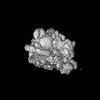 37007MC 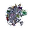 8xz3C M: map data used to model this data C: citing same article ( |
|---|---|
| Similar structure data |
-Links
 PDBj
PDBj


- Assembly
| Deposited unit | 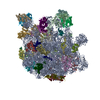
|
|---|---|
| 1 |
|
-Components
+50S ribosomal protein ... , 32 types, 32 molecules 3CDEFGHIJKLMNOPQRSTUVWXYZabcdefg
-RNA chain , 2 types, 2 molecules AB
| #2: RNA chain | Mass: 1012140.938 Da / Num. of mol.: 1 / Source method: isolated from a natural source Source: (natural) Mycolicibacterium smegmatis MC2 155 (bacteria)References: GenBank: 118168627 |
|---|---|
| #3: RNA chain | Mass: 38061.816 Da / Num. of mol.: 1 / Source method: isolated from a natural source Source: (natural) Mycolicibacterium smegmatis MC2 155 (bacteria)References: GenBank: 118168627 |
-Protein / Non-polymers , 2 types, 2 molecules h
| #35: Protein | Mass: 52163.805 Da / Num. of mol.: 1 Source method: isolated from a genetically manipulated source Source: (gene. exp.) Mycolicibacterium smegmatis MC2 155 (bacteria)Gene: hflX / Production host: |
|---|---|
| #36: Chemical | ChemComp-GNP /  Mass: 522.196 Da / Num. of mol.: 1 / Source method: obtained synthetically / Formula: C10H17N6O13P3 / Feature type: SUBJECT OF INVESTIGATION Mass: 522.196 Da / Num. of mol.: 1 / Source method: obtained synthetically / Formula: C10H17N6O13P3 / Feature type: SUBJECT OF INVESTIGATIONComment: GppNHp, GMPPNP, energy-carrying molecule analogue*YM |
-Details
| Has ligand of interest | Y |
|---|
-Experimental details
-Experiment
| Experiment | Method: ELECTRON MICROSCOPY |
|---|---|
| EM experiment | Aggregation state: PARTICLE / 3D reconstruction method: single particle reconstruction |
- Sample preparation
| Component |
| ||||||||||||||||||||||||
|---|---|---|---|---|---|---|---|---|---|---|---|---|---|---|---|---|---|---|---|---|---|---|---|---|---|
| Source (natural) |
| ||||||||||||||||||||||||
| Source (recombinant) |
| ||||||||||||||||||||||||
| Buffer solution | pH: 7.5 | ||||||||||||||||||||||||
| Specimen | Embedding applied: NO / Shadowing applied: NO / Staining applied: NO / Vitrification applied: YES | ||||||||||||||||||||||||
| Vitrification | Cryogen name: ETHANE |
- Electron microscopy imaging
| Experimental equipment |  Model: Titan Krios / Image courtesy: FEI Company |
|---|---|
| Microscopy | Model: FEI TITAN KRIOS |
| Electron gun | Electron source:  FIELD EMISSION GUN / Accelerating voltage: 300 kV / Illumination mode: FLOOD BEAM FIELD EMISSION GUN / Accelerating voltage: 300 kV / Illumination mode: FLOOD BEAM |
| Electron lens | Mode: BRIGHT FIELD / Nominal defocus max: 3300 nm / Nominal defocus min: 1800 nm |
| Image recording | Electron dose: 28.26 e/Å2 / Film or detector model: FEI FALCON III (4k x 4k) |
- Processing
| EM software |
| ||||||||||||||||||||
|---|---|---|---|---|---|---|---|---|---|---|---|---|---|---|---|---|---|---|---|---|---|
| CTF correction | Type: PHASE FLIPPING AND AMPLITUDE CORRECTION | ||||||||||||||||||||
| 3D reconstruction | Resolution: 3.3 Å / Resolution method: FSC 0.143 CUT-OFF / Num. of particles: 121211 / Symmetry type: POINT |
