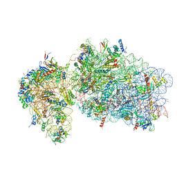
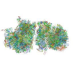
3J6Y
 
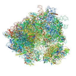 | | S. cerevisiae 80S ribosome bound with Taura syndrome virus (TSV) IRES, 2 degree rotation (Class I) | | 分子名称: | 18S ribosomal RNA, 25S ribosomal RNA, 40S ribosomal protein S0, ... | | 著者 | Koh, C.S, Brilot, A.F, Grigorieff, N, Korostelev, A.A. | | 登録日 | 2014-04-16 | | 公開日 | 2014-06-11 | | 最終更新日 | 2024-02-21 | | 実験手法 | ELECTRON MICROSCOPY (6.1 Å) | | 主引用文献 | Taura syndrome virus IRES initiates translation by binding its tRNA-mRNA-like structural element in the ribosomal decoding center.
Proc.Natl.Acad.Sci.USA, 111, 2014
|
|
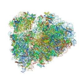
6R6P
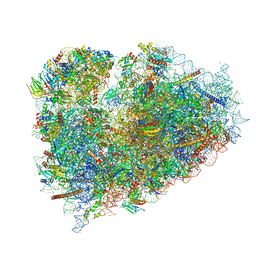 | | Structure of XBP1u-paused ribosome nascent chain complex (rotated state) | | 分子名称: | 18S rRNA, 28S ribosomal RNA, 40S ribosomal protein S12, ... | | 著者 | Shanmuganathan, V, Cheng, J, Berninghausen, O, Beckmann, R. | | 登録日 | 2019-03-27 | | 公開日 | 2019-07-10 | | 最終更新日 | 2024-05-22 | | 実験手法 | ELECTRON MICROSCOPY (3.1 Å) | | 主引用文献 | Structural and mutational analysis of the ribosome-arresting human XBP1u.
Elife, 8, 2019
|
|
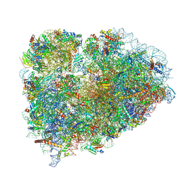
6R6G
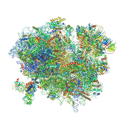 | | Structure of XBP1u-paused ribosome nascent chain complex with SRP. | | 分子名称: | 18S ribosomal RNA, 28S ribosomal RNA, 40S ribosomal protein S12, ... | | 著者 | Shanmuganathan, V, Cheng, J, Braunger, K, Berninghausen, O, Beatrix, B, Beckmann, R. | | 登録日 | 2019-03-27 | | 公開日 | 2019-07-10 | | 最終更新日 | 2019-10-30 | | 実験手法 | ELECTRON MICROSCOPY (3.7 Å) | | 主引用文献 | Structural and mutational analysis of the ribosome-arresting human XBP1u.
Elife, 8, 2019
|
|
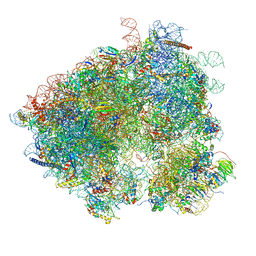
6R7Q
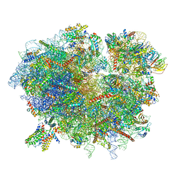 | | Structure of XBP1u-paused ribosome nascent chain complex with Sec61. | | 分子名称: | 18S ribosomal RNA, 28S ribosomal RNA, 40S ribosomal protein S12, ... | | 著者 | Shanmuganathan, V, Cheng, J, Braunger, K, Berninghausen, O, Beatrix, B, Beckmann, R. | | 登録日 | 2019-03-29 | | 公開日 | 2019-07-10 | | 最終更新日 | 2024-05-22 | | 実験手法 | ELECTRON MICROSCOPY (3.9 Å) | | 主引用文献 | Structural and mutational analysis of the ribosome-arresting human XBP1u.
Elife, 8, 2019
|
|
6RBE
 | | State 2 of yeast Tsr1-TAP Rps20-Deltaloop pre-40S particles | | 分子名称: | 18S ribosomal RNA, 40S ribosomal protein S0-A, 40S ribosomal protein S1-A, ... | | 著者 | Shayan, R, Mitterer, V, Ferreira-Cerca, S, Murat, G, Enne, T, Rinaldi, D, Weigl, S, Omanic, H, Gleizes, P.E, Kressler, D, Pertschy, B, Plisson-Chastang, C. | | 登録日 | 2019-04-10 | | 公開日 | 2019-06-26 | | 最終更新日 | 2024-05-22 | | 実験手法 | ELECTRON MICROSCOPY (3.8 Å) | | 主引用文献 | Conformational proofreading of distant 40S ribosomal subunit maturation events by a long-range communication mechanism.
Nat Commun, 10, 2019
|
|
6SGC
 | | Rabbit 80S ribosome stalled on a poly(A) tail | | 分子名称: | 18S ribosomal RNA, 28S ribosomal RNA, 40S ribosomal protein S12, ... | | 著者 | Chandrasekaran, V, Juszkiewicz, S, Choi, J, Puglisi, J.D, Brown, A, Shao, S, Ramakrishnan, V, Hegde, R.S. | | 登録日 | 2019-08-03 | | 公開日 | 2019-12-04 | | 最終更新日 | 2022-03-30 | | 実験手法 | ELECTRON MICROSCOPY (2.8 Å) | | 主引用文献 | Mechanism of ribosome stalling during translation of a poly(A) tail.
Nat.Struct.Mol.Biol., 26, 2019
|
|
3JAH
 | | Structure of a mammalian ribosomal termination complex with ABCE1, eRF1(AAQ), and the UAG stop codon | | 分子名称: | 18S ribosomal RNA, 28S ribosomal RNA, 5.8S ribosomal RNA, ... | | 著者 | Brown, A, Shao, S, Murray, J, Hegde, R.S, Ramakrishnan, V. | | 登録日 | 2015-06-10 | | 公開日 | 2015-08-12 | | 最終更新日 | 2018-07-18 | | 実験手法 | ELECTRON MICROSCOPY (3.45 Å) | | 主引用文献 | Structural basis for stop codon recognition in eukaryotes.
Nature, 524, 2015
|
|
3J7P
 | | Structure of the 80S mammalian ribosome bound to eEF2 | | 分子名称: | 18S ribosomal RNA, 28S ribosomal RNA, 5.8S ribosomal RNA, ... | | 著者 | Voorhees, R.M, Fernandez, I.S, Scheres, S.H.W, Hegde, R.S. | | 登録日 | 2014-08-01 | | 公開日 | 2014-09-03 | | 最終更新日 | 2018-07-18 | | 実験手法 | ELECTRON MICROSCOPY (3.5 Å) | | 主引用文献 | Structure of the Mammalian ribosome-sec61 complex to 3.4 a resolution.
Cell(Cambridge,Mass.), 157, 2014
|
|
3JAM
 | | CryoEM structure of 40S-eIF1A-eIF1 complex from yeast | | 分子名称: | 18S rRNA, MAGNESIUM ION, RACK1, ... | | 著者 | Llacer, J.L, Hussain, T, Ramakrishnan, V. | | 登録日 | 2015-06-17 | | 公開日 | 2015-08-12 | | 最終更新日 | 2024-02-21 | | 実験手法 | ELECTRON MICROSCOPY (3.46 Å) | | 主引用文献 | Conformational Differences between Open and Closed States of the Eukaryotic Translation Initiation Complex.
Mol.Cell, 59, 2015
|
|
6R5Q
 | | Structure of XBP1u-paused ribosome nascent chain complex (post-state) | | 分子名称: | 18S ribosomal RNA, 28S ribosomal RNA, 40S ribosomal protein S12, ... | | 著者 | Shanmuganathan, V, Cheng, J, Berninghausen, O, Beckmann, R. | | 登録日 | 2019-03-25 | | 公開日 | 2019-07-10 | | 最終更新日 | 2019-10-30 | | 実験手法 | ELECTRON MICROSCOPY (3 Å) | | 主引用文献 | Structural and mutational analysis of the ribosome-arresting human XBP1u.
Elife, 8, 2019
|
|
3J6X
 | | S. cerevisiae 80S ribosome bound with Taura syndrome virus (TSV) IRES, 5 degree rotation (Class II) | | 分子名称: | 18S ribosomal RNA, 25S ribosomal RNA, 40S ribosomal protein S0, ... | | 著者 | Koh, C.S, Brilot, A.F, Grigorieff, N, Korostelev, A.A. | | 登録日 | 2014-04-16 | | 公開日 | 2014-06-11 | | 最終更新日 | 2024-02-21 | | 実験手法 | ELECTRON MICROSCOPY (6.1 Å) | | 主引用文献 | Taura syndrome virus IRES initiates translation by binding its tRNA-mRNA-like structural element in the ribosomal decoding center.
Proc.Natl.Acad.Sci.USA, 111, 2014
|
|
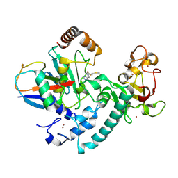
3K9O
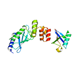 | | The crystal structure of E2-25K and UBB+1 complex | | 分子名称: | Ubiquitin, Ubiquitin-conjugating enzyme E2 K | | 著者 | Kang, G.B, Ko, S, Song, S.M, Lee, W, Eom, S.H. | | 登録日 | 2009-10-16 | | 公開日 | 2010-09-08 | | 最終更新日 | 2024-03-20 | | 実験手法 | X-RAY DIFFRACTION (1.8 Å) | | 主引用文献 | Structural Basis of E2-25K/UBB+1 Interaction for Neurotoxicity of Alzheimer Disease by Proteasome Inhibition
To be Published
|
|
3J80
 | | CryoEM structure of 40S-eIF1-eIF1A preinitiation complex | | 分子名称: | 18S rRNA, MAGNESIUM ION, RACK1, ... | | 著者 | Hussain, T, Llacer, J.L, Fernandez, I.S, Savva, C.G, Ramakrishnan, V. | | 登録日 | 2014-08-28 | | 公開日 | 2014-11-05 | | 最終更新日 | 2024-02-21 | | 実験手法 | ELECTRON MICROSCOPY (3.75 Å) | | 主引用文献 | Structural changes enable start codon recognition by the eukaryotic translation initiation complex.
Cell(Cambridge,Mass.), 159, 2014
|
|
3JAG
 | | Structure of a mammalian ribosomal termination complex with ABCE1, eRF1(AAQ), and the UAA stop codon | | 分子名称: | 18S ribosomal RNA, 28S ribosomal RNA, 5.8S ribosomal RNA, ... | | 著者 | Brown, A, Shao, S, Murray, J, Hegde, R.S, Ramakrishnan, V. | | 登録日 | 2015-06-10 | | 公開日 | 2015-08-12 | | 最終更新日 | 2018-07-18 | | 実験手法 | ELECTRON MICROSCOPY (3.65 Å) | | 主引用文献 | Structural basis for stop codon recognition in eukaryotes.
Nature, 524, 2015
|
|
3K9P
 | | The crystal structure of E2-25K and ubiquitin complex | | 分子名称: | Ubiquitin, Ubiquitin-conjugating enzyme E2 K | | 著者 | Kang, G.B, Ko, S, Song, S.M, Lee, W, Eom, S.H. | | 登録日 | 2009-10-16 | | 公開日 | 2010-09-08 | | 最終更新日 | 2024-03-20 | | 実験手法 | X-RAY DIFFRACTION (2.8 Å) | | 主引用文献 | Structural basis of E2-25K/UBB+1 interaction leading to proteasome inhibition and neurotoxicity
J.Biol.Chem., 285, 2010
|
|
3J79
 | | Cryo-EM structure of the Plasmodium falciparum 80S ribosome bound to the anti-protozoan drug emetine, large subunit | | 分子名称: | 28S ribosomal RNA, 5.8S ribosomal RNA, 5S ribosomal RNA, ... | | 著者 | Wong, W, Bai, X.C, Brown, A, Fernandez, I.S, Hanssen, E, Condron, M, Tan, Y.H, Baum, J, Scheres, S.H.W. | | 登録日 | 2014-06-02 | | 公開日 | 2014-07-16 | | 最終更新日 | 2024-02-21 | | 実験手法 | ELECTRON MICROSCOPY (3.2 Å) | | 主引用文献 | Cryo-EM structure of the Plasmodium falciparum 80S ribosome bound to the anti-protozoan drug emetine.
Elife, 3, 2014
|
|
3JW0
 | | E2~Ubiquitin-HECT | | 分子名称: | E3 ubiquitin-protein ligase NEDD4-like, Ubiquitin, Ubiquitin-conjugating enzyme E2 D2 | | 著者 | Kamadurai, H.B, Schulman, B.A. | | 登録日 | 2009-09-17 | | 公開日 | 2010-01-12 | | 最終更新日 | 2021-10-13 | | 実験手法 | X-RAY DIFFRACTION (3.1 Å) | | 主引用文献 | Insights into ubiquitin transfer cascades from a structure of a UbcH5B approximately ubiquitin-HECT(NEDD4L) complex.
Mol.Cell, 36, 2009
|
|
3J7Q
 | | Structure of the idle mammalian ribosome-Sec61 complex | | 分子名称: | 28S ribosomal RNA, 5.8S ribosomal RNA, 5S ribosomal RNA, ... | | 著者 | Voorhees, R.M, Fernandez, I.S, Scheres, S.H.W, Hegde, R.S. | | 登録日 | 2014-08-01 | | 公開日 | 2014-09-03 | | 最終更新日 | 2019-10-30 | | 実験手法 | ELECTRON MICROSCOPY (3.5 Å) | | 主引用文献 | Structure of the Mammalian ribosome-sec61 complex to 3.4 a resolution.
Cell(Cambridge,Mass.), 157, 2014
|
|
3JAP
 | | Structure of a partial yeast 48S preinitiation complex in closed conformation | | 分子名称: | 18S rRNA, MAGNESIUM ION, METHIONINE, ... | | 著者 | Llacer, J.L, Hussain, T, Ramakrishnan, V. | | 登録日 | 2015-06-18 | | 公開日 | 2015-08-12 | | 最終更新日 | 2024-02-21 | | 実験手法 | ELECTRON MICROSCOPY (4.9 Å) | | 主引用文献 | Conformational Differences between Open and Closed States of the Eukaryotic Translation Initiation Complex.
Mol.Cell, 59, 2015
|
|
3JAI
 | | Structure of a mammalian ribosomal termination complex with ABCE1, eRF1(AAQ), and the UGA stop codon | | 分子名称: | 18S ribosomal RNA, 28S ribosomal RNA, 5.8S ribosomal RNA, ... | | 著者 | Brown, A, Shao, S, Murray, J, Hegde, R.S, Ramakrishnan, V. | | 登録日 | 2015-06-10 | | 公開日 | 2015-08-12 | | 最終更新日 | 2018-07-18 | | 実験手法 | ELECTRON MICROSCOPY (3.65 Å) | | 主引用文献 | Structural basis for stop codon recognition in eukaryotes.
Nature, 524, 2015
|
|
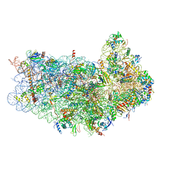
6T7I
 | | Structure of yeast 80S ribosome stalled on the CGA-CGA inhibitory codon combination. | | 分子名称: | 18S ribosomal RNA, 25S ribosomal RNA, 40S ribosomal protein S0-A, ... | | 著者 | Tesina, P, Buschauer, R, Cheng, J, Berninghausen, O, Becker, R, Beckmann, R. | | 登録日 | 2019-10-22 | | 公開日 | 2019-12-25 | | 最終更新日 | 2024-05-22 | | 実験手法 | ELECTRON MICROSCOPY (3.2 Å) | | 主引用文献 | Molecular mechanism of translational stalling by inhibitory codon combinations and poly(A) tracts.
Embo J., 39, 2020
|
|
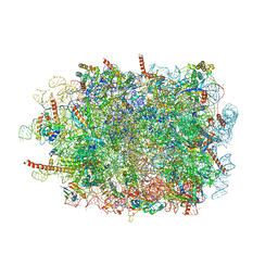
6RI5
 | |
3J78
 | | Structures of yeast 80S ribosome-tRNA complexes in the rotated and non-rotated conformations (Class I - non-rotated ribosome with 2 tRNAs) | | 分子名称: | 18S ribosomal RNA, 25S ribosomal RNA, 40S ribosomal protein S0, ... | | 著者 | Svidritskiy, E, Brilot, A.F, Koh, C.S, Grigorieff, N, Korostelev, A.A. | | 登録日 | 2014-05-29 | | 公開日 | 2014-08-06 | | 最終更新日 | 2024-02-21 | | 実験手法 | ELECTRON MICROSCOPY (6.3 Å) | | 主引用文献 | Structures of Yeast 80S Ribosome-tRNA Complexes in the Rotated and Nonrotated Conformations.
Structure, 22, 2014
|
|
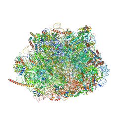
6T83
 | | Structure of yeast disome (di-ribosome) stalled on poly(A) tract. | | 分子名称: | 18S rRNA, 25S rRNA, 40S ribosomal protein S0-A, ... | | 著者 | Tesina, P, Buschauer, R, Cheng, J, Berninghausen, O, Becker, R, Beckmann, R. | | 登録日 | 2019-10-24 | | 公開日 | 2019-12-25 | | 最終更新日 | 2020-02-12 | | 実験手法 | ELECTRON MICROSCOPY (4 Å) | | 主引用文献 | Molecular mechanism of translational stalling by inhibitory codon combinations and poly(A) tracts.
Embo J., 39, 2020
|
|
6T7F
 | | RCR E3 ligase E2-Ubiquitin transthiolation intermediate | | 分子名称: | 3,3-bis(sulfanyl)-~{N}-(1~{H}-1,2,3-triazol-4-ylmethyl)propanamide, E3 ubiquitin-protein ligase MYCBP2, Polyubiquitin-C, ... | | 著者 | Mabbitt, P.D, Virdee, S. | | 登録日 | 2019-10-21 | | 公開日 | 2020-08-05 | | 最終更新日 | 2024-01-24 | | 実験手法 | X-RAY DIFFRACTION (2.58 Å) | | 主引用文献 | Structural basis for RING-Cys-Relay E3 ligase activity and its role in axon integrity.
Nat.Chem.Biol., 16, 2020
|
|


