 Movie
Movie Controller

[English] 日本語
 Yorodumi
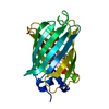
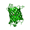
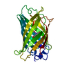
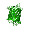
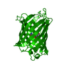
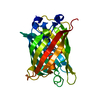
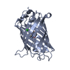
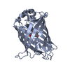
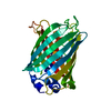
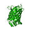
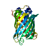
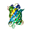
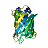
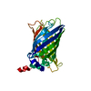
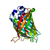
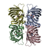
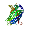
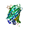
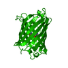
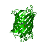
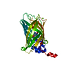
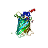
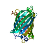
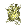
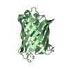
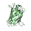
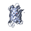
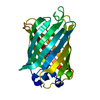
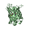
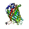
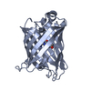
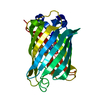
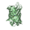
- PDB-2wsn: Structure of Enhanced Cyan Fluorescent Protein at physiological pH -
+ Open data

- Basic information
| Entry | Database: PDB / ID: 2wsn | ||||||
|---|---|---|---|---|---|---|---|
| Title | Structure of Enhanced Cyan Fluorescent Protein at physiological pH | ||||||
 Components Components | GREEN FLUORESCENT PROTEIN | ||||||
 Keywords Keywords | FLUORESCENT PROTEIN / CHROMOPHORE / LUMINESCENCE / PHOTOPROTEIN | ||||||
| Function / homology |  Function and homology information Function and homology information | ||||||
| Biological species |   AEQUOREA VICTORIA (jellyfish) AEQUOREA VICTORIA (jellyfish) | ||||||
| Method |  X-RAY DIFFRACTION / SYNCHROTRON / MOLECULAR REPLACEMENT / Resolution: 1.37 Å X-RAY DIFFRACTION / SYNCHROTRON / MOLECULAR REPLACEMENT / Resolution: 1.37 Å | ||||||
 Authors Authors | Lelimousin, M. / Noirclerc-Savoye, M. / Lazareno-Saez, C. / Paetzold, B. / Le Vot, S. / Chazal, R. / Macheboeuf, P. / Field, M.J. / Bourgeois, D. / Royant, A. | ||||||
 Citation Citation | Journal: Biochemistry / Year: 2009 Title: Intrinsic Dynamics in Ecfp and Cerulean Control Fluorescence Quantum Yield. Authors: Lelimousin, M. / Noirclerc-Savoye, M. / Lazareno-Saez, C. / Paetzold, B. / Le Vot, S. / Chazal, R. / Macheboeuf, P. / Field, M.J. / Bourgeois, D. / Royant, A. #1: Journal: J.Mol.Biol. / Year: 2003Title: Expansion of the Genetic Code Enables Design of a Novel "Gold" Class of Green Fluorescent Proteins. Authors: Bae, J.H. / Rubini, M. / Jung, G. / Wiegand, G. / Seifert, M.H.J. / Azim, M.K. / Kim, J.S. / Zumbusch, A. / Holak, T.A. / Moroder, L. / Huber, R. / Budisa, N. | ||||||
| History |
|
- Structure visualization
| Structure viewer | Molecule: MolmilJmol/JSmol |
|---|
- Downloads & links
-Download
| PDBx/mmCIF format | 2wsn.cif.gz | 118.2 KB | Display | PDBx/mmCIF format |
|---|---|---|---|---|
| PDB format | pdb2wsn.ent.gz | 92.7 KB | Display | PDB format |
| PDBx/mmJSON format | 2wsn.json.gz | Tree view | PDBx/mmJSON format | |
| Others |  Other downloads Other downloads |
-Validation report
| Arichive directory | https://data.pdbj.org/pub/pdb/validation_reports/ws/2wsnftp://data.pdbj.org/pub/pdb/validation_reports/ws/2wsn | HTTPS FTP |
|---|
-Related structure data
| Related structure data | 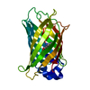 2wsoC  1oxeS C: citing same article ( S: Starting model for refinement |
|---|---|
| Similar structure data |


-Links
 PDBj
PDBj

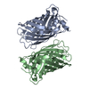
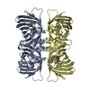
- Assembly
| Deposited unit | 
| ||||||||
|---|---|---|---|---|---|---|---|---|---|
| 1 |
| ||||||||
| Unit cell |
|
-Components
| #1: Protein | Mass: 26920.393 Da / Num. of mol.: 1 / Fragment: RESIDUES 2-238 Source method: isolated from a genetically manipulated source Details: CHROMOPHORE RESULTING FROM AUTOCATALYTIC CYCLIZATION OF CONSECUTIVE AMINO ACID RESIDUES THR65, TRP66 AND GLY67. Source: (gene. exp.) AEQUOREA VICTORIA (jellyfish) / Production host:  |
|---|---|
| #2: Water | ChemComp-HOH /  Mass: 18.015 Da / Num. of mol.: 224 / Source method: isolated from a natural source / Formula: H2O Mass: 18.015 Da / Num. of mol.: 224 / Source method: isolated from a natural source / Formula: H2O |
| Has protein modification | Y |
| Sequence details | GFP VARIANT |
-Experimental details
-Experiment
| Experiment | Method: X-RAY DIFFRACTION / Number of used crystals: 1 |
|---|
- Sample preparation
| Crystal | Density Matthews: 2 Å3/Da / Density % sol: 40 % / Description: NONE |
|---|---|
| Crystal grow | pH: 7.5 / Details: 19.5% PEG8000, 4% GLYCEROL, 1M HEPES PH 7.5 |
-Data collection
| Diffraction | Mean temperature: 100 K |
|---|---|
| Diffraction source | Source: SYNCHROTRON / Site: ESRF  / Beamline: ID14-2 / Wavelength: 0.933 / Beamline: ID14-2 / Wavelength: 0.933 |
| Detector | Type: ADSC CCD / Detector: CCD / Date: Jun 13, 2005 / Details: TOROIDAL MIRROR |
| Radiation | Monochromator: DIAMOND / Protocol: SINGLE WAVELENGTH / Monochromatic (M) / Laue (L): M / Scattering type: x-ray |
| Radiation wavelength | Wavelength: 0.933 Å / Relative weight: 1 |
| Reflection | Resolution: 1.37→40 Å / Num. obs: 45997 / % possible obs: 94.9 % / Observed criterion σ(I): 2 / Redundancy: 4.7 % / Biso Wilson estimate: 17.4 Å2 / Rmerge(I) obs: 0.05 / Net I/σ(I): 22.3 |
| Reflection shell | Resolution: 1.37→1.42 Å / Redundancy: 3.7 % / Rmerge(I) obs: 0.41 / Mean I/σ(I) obs: 3.3 / % possible all: 70.4 |
- Processing
| Software |
| ||||||||||||||||||||||||||||||||||||||||||||||||||||||||||||||||||||||||||||||||||||||||||||||||||||||||||||||||||||||||||||||||||||||||||||||||||||||||||||||||||||||||||||||||||||||
|---|---|---|---|---|---|---|---|---|---|---|---|---|---|---|---|---|---|---|---|---|---|---|---|---|---|---|---|---|---|---|---|---|---|---|---|---|---|---|---|---|---|---|---|---|---|---|---|---|---|---|---|---|---|---|---|---|---|---|---|---|---|---|---|---|---|---|---|---|---|---|---|---|---|---|---|---|---|---|---|---|---|---|---|---|---|---|---|---|---|---|---|---|---|---|---|---|---|---|---|---|---|---|---|---|---|---|---|---|---|---|---|---|---|---|---|---|---|---|---|---|---|---|---|---|---|---|---|---|---|---|---|---|---|---|---|---|---|---|---|---|---|---|---|---|---|---|---|---|---|---|---|---|---|---|---|---|---|---|---|---|---|---|---|---|---|---|---|---|---|---|---|---|---|---|---|---|---|---|---|---|---|---|---|
| Refinement | Method to determine structure: MOLECULAR REPLACEMENT Starting model: PDB ENTRY 1OXE Resolution: 1.37→35.29 Å / Cor.coef. Fo:Fc: 0.971 / Cor.coef. Fo:Fc free: 0.958 / SU B: 1.976 / SU ML: 0.035 / Cross valid method: THROUGHOUT / ESU R: 0.06 / ESU R Free: 0.059 / Stereochemistry target values: MAXIMUM LIKELIHOOD Details: HYDROGENS HAVE BEEN ADDED IN THE RIDING POSITIONS. U VALUES REFINED INDIVIDUALLY
| ||||||||||||||||||||||||||||||||||||||||||||||||||||||||||||||||||||||||||||||||||||||||||||||||||||||||||||||||||||||||||||||||||||||||||||||||||||||||||||||||||||||||||||||||||||||
| Solvent computation | Ion probe radii: 0.8 Å / Shrinkage radii: 0.8 Å / VDW probe radii: 1.2 Å / Solvent model: MASK | ||||||||||||||||||||||||||||||||||||||||||||||||||||||||||||||||||||||||||||||||||||||||||||||||||||||||||||||||||||||||||||||||||||||||||||||||||||||||||||||||||||||||||||||||||||||
| Displacement parameters | Biso mean: 9.8 Å2
| ||||||||||||||||||||||||||||||||||||||||||||||||||||||||||||||||||||||||||||||||||||||||||||||||||||||||||||||||||||||||||||||||||||||||||||||||||||||||||||||||||||||||||||||||||||||
| Refinement step | Cycle: LAST / Resolution: 1.37→35.29 Å
| ||||||||||||||||||||||||||||||||||||||||||||||||||||||||||||||||||||||||||||||||||||||||||||||||||||||||||||||||||||||||||||||||||||||||||||||||||||||||||||||||||||||||||||||||||||||
| Refine LS restraints |
|