 Movie
Movie Controller


+ Open data

- Basic information
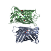
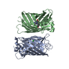
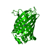
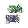
| Entry | Database: PDB / ID: 1gfl | ||||||
|---|---|---|---|---|---|---|---|
| Title | STRUCTURE OF GREEN FLUORESCENT PROTEIN | ||||||
 Components Components | GREEN FLUORESCENT PROTEIN | ||||||
 Keywords Keywords | FLUORESCENT PROTEIN / FLUOROPHORE GREEN FLUORESCENT PROTEIN / LUMINESCENCE | ||||||
| Function / homology |  Function and homology information Function and homology information | ||||||
| Biological species |   Aequorea victoria (jellyfish) Aequorea victoria (jellyfish) | ||||||
| Method |  X-RAY DIFFRACTION / Resolution: 1.9 Å X-RAY DIFFRACTION / Resolution: 1.9 Å | ||||||
 Authors Authors | Yang, F. / Moss, L.G. / Phillips Jr., G.N. | ||||||
 Citation Citation | Journal: Nat.Biotechnol. / Year: 1996 Title: The molecular structure of green fluorescent protein. Authors: Yang, F. / Moss, L.G. / Phillips Jr., G.N. #1: Journal: Trends Biochem.Sci. / Year: 1995Title: Understanding, Improving and Using Green Fluorescent Proteins Authors: Cubitt, A.B. / Heim, R. / Adams, S.R. / Boyd, A.E. / Gross, L.A. / Tsien, R.Y. | ||||||
| History |
|
- Structure visualization
| Structure viewer | Molecule: MolmilJmol/JSmol |
|---|
- Downloads & links
-Download
| PDBx/mmCIF format | 1gfl.cif.gz | 104.7 KB | Display | PDBx/mmCIF format |
|---|---|---|---|---|
| PDB format | pdb1gfl.ent.gz | 81 KB | Display | PDB format |
| PDBx/mmJSON format | 1gfl.json.gz | Tree view | PDBx/mmJSON format | |
| Others |  Other downloads Other downloads |
-Validation report
| Arichive directory | https://data.pdbj.org/pub/pdb/validation_reports/gf/1gflftp://data.pdbj.org/pub/pdb/validation_reports/gf/1gfl | HTTPS FTP |
|---|
-Related structure data
| Similar structure data |
|---|


-Links
 PDBj
PDBj

- Assembly
| Deposited unit | 
| ||||||||
|---|---|---|---|---|---|---|---|---|---|
| 1 | 
| ||||||||
| 2 | 
| ||||||||
| Unit cell |
| ||||||||
| Noncrystallographic symmetry (NCS) | NCS oper: (Code: given Matrix: (-0.950278, 0.287772, 0.118992), Vector: |
-Components
| #1: Protein | Mass: 26891.271 Da / Num. of mol.: 2 / Mutation: Q80R Source method: isolated from a genetically manipulated source Source: (gene. exp.) Aequorea victoria (jellyfish) / Plasmid: PTU58 / Production host:  #2: Water | ChemComp-HOH / |  Mass: 18.015 Da / Num. of mol.: 300 / Source method: isolated from a natural source / Formula: H2O Mass: 18.015 Da / Num. of mol.: 300 / Source method: isolated from a natural source / Formula: H2OHas protein modification | Y | Sequence details | THE FLUOROPHORE IS FORMED BY SER 65, TYR 66 AND GLY 67. THE CARBONYL CARBON OF TYR 66 IS BONDED TO ...THE FLUOROPHOR | |
|---|
-Experimental details
-Experiment
| Experiment | Method: X-RAY DIFFRACTION |
|---|
- Sample preparation
| Crystal | Density Matthews: 2.22 Å3/Da / Density % sol: 44.48 % |
|---|---|
| Crystal grow | pH: 7 / Details: FREE TEXT GOES HERE., pH 7.0 |
-Data collection
| Diffraction | Mean temperature: 299 K |
|---|---|
| Detector | Type: RIGAKU / Detector: IMAGE PLATE / Date: Mar 25, 1995 |
| Radiation | Scattering type: x-ray |
| Radiation wavelength | Relative weight: 1 |
| Reflection | Resolution: 1.9→30 Å / Num. obs: 38472 / % possible obs: 99.5 % / Observed criterion σ(I): 0 / Redundancy: 5 % / Rmerge(I) obs: 0.077 |
- Processing
| Software |
| ||||||||||||||||||||||||||||||||||||||||||||||||||||||||||||
|---|---|---|---|---|---|---|---|---|---|---|---|---|---|---|---|---|---|---|---|---|---|---|---|---|---|---|---|---|---|---|---|---|---|---|---|---|---|---|---|---|---|---|---|---|---|---|---|---|---|---|---|---|---|---|---|---|---|---|---|---|---|
| Refinement | Resolution: 1.9→10 Å / σ(F): 0
| ||||||||||||||||||||||||||||||||||||||||||||||||||||||||||||
| Refinement step | Cycle: LAST / Resolution: 1.9→10 Å
| ||||||||||||||||||||||||||||||||||||||||||||||||||||||||||||
| Refine LS restraints |
|
